14
views
0
recommends
+1 Recommend
0 collections
    0
    shares
      • Record: found
      • Abstract: found
      • Article: found
      Is Open Access

      Celastrol Induces Necroptosis and Ameliorates Inflammation via Targeting Biglycan in Human Gastric Carcinoma

      research-article

      Read this article at

          There is no author summary for this article yet. Authors can add summaries to their articles on ScienceOpen to make them more accessible to a non-specialist audience.

          Abstract

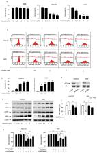
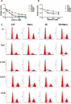
          Celastrol, a triterpene isolated from the root of traditional Chinese medicine Thunder of God Vine, possesses anti-cancer and anti-inflammatory activity to treat rheumatoid disease or as health product. Necroptosis is considered as a new approach to overcome chemotherapeutics resistance. However, whether celastrol exerts necroptosis leading to gastric cancer cell death is still unclear. Here, for the first time we showed that celastrol induced necroptosis in HGC27 and AGS gastric cancer cell lines. More importantly, celastrol down-regulated biglycan (BGN) protein, which is critical for gastric cancer migration and invasion. Furthermore, celastrol activated receptor-interacting protein 1 and 3 (RIP1 and RIP3) and subsequently promoted the translation of mixed-lineage kinase domain-like (MLKL) from cytoplasm to plasma membrane, leading to necroptosis of gastric cancer cell, which was blocked by over-expression BGN. In addition, celastrol suppressed the release of pro-inflammatory cytokines TNF-α and IL-8 in HGC27 and AGS cells, which was reversed by over-expression BGN. Taken together, we identified celastrol as a necroptosis inducer, activated RIP1/RIP3/MLKL pathway and suppressed the level of pro-inflammatory cytokines by down-regulating BGN in HGC-27 and AGS cells, which supported the feasibility of celastrol in gastric cancer therapy.

          Related collections

          Most cited references40

          • Record: found
          • Abstract: not found
          • Article: not found

          An Inflammatory Perspective on Necroptosis

            Bookmark
            • Record: found
            • Abstract: found
            • Article: not found

            Targeted disruption of the biglycan gene leads to an osteoporosis-like phenotype in mice.

            The resilience and strength of bone is due to the orderly mineralization of a specialized extracellular matrix (ECM) composed of type I collagen (90%) and a host of non-collagenous proteins that are, in general, also found in other tissues. Biglycan (encoded by the gene Bgn) is an ECM proteoglycan that is enriched in bone and other non-skeletal connective tissues. In vitro studies indicate that Bgn may function in connective tissue metabolism by binding to collagen fibrils and TGF-beta (refs 5,6), and may promote neuronal survival. To study the role of Bgn in vivo, we generated Bgn-deficient mice. Although apparently normal at birth, these mice display a phenotype characterized by a reduced growth rate and decreased bone mass due to the absence of Bgn. To our knowledge, this is the first report in which deficiency of a non-collagenous ECM protein leads to a skeletal phenotype that is marked by low bone mass that becomes more obvious with age. These mice may serve as an animal model to study the role of ECM proteins in osteoporosis.
              Bookmark
              • Record: found
              • Abstract: found
              • Article: found
              Is Open Access

              The anti-tumor effect of shikonin on osteosarcoma by inducing RIP1 and RIP3 dependent necroptosis

              Background Osteosarcoma is the most frequent primary malignant bone tumor, notorious for its lung metastasis. Shikonin, an effective constituent extracted from Chinese medicinal herb, was demonstrated to induce necroptosis in some cancers. Methods MTT assay was performed to detect cell survival rate in vitro. Flow cytometry was used to analyze cell cycle and cell death. Western blot was performed to determine the expression levels of RIP1, RIP3, caspase-3, caspase-6 and PARP. The tibial primary and lung metastatic osteosarcoma models were used to evaluate the anti-tumor effect of shikonin in vivo. Results The cell survival rate was decreased in a dose and time dependent manner when treated with shikonin. No major change in cell cycle was observed after shikonin treatment. The cell death induced by shikonin could be mostly rescued by specific necroptosis inhibitor necrostatin-1, but not by general caspase inhibitor Z-VAD-FMK. The number of necrotic cells caused by shikonin was decreased after being pretreated with Nec-1 detected by flow cytometry in K7 cells. After 8-hour treatment of shikonin, the expression levels of RIP1 and RIP3 were increased while caspase-3, caspase-6 and PARP were not activated in K7 and U2OS cells determined by Western blot. Size of primary tumor and lung metastasis in shikonin treated group were significantly reduced. The protein levels of RIP1 and RIP3 in primary tumor tissues were increased by shikonin. The overall survival of lung metastatic models was longer compared with control group (p < 0.001). Conclusions Shikonin had prompt but profound anti-tumor effect on both primary and metastatic osteosarcoma, probably by inducing RIP1 and RIP3 dependent necroptosis. Shikonin would be a potential anti-tumor agent on the treatment of primary and metastatic osteosarcoma.
                Bookmark

                Author and article information

                Journal
                Int J Mol Sci
                Int J Mol Sci
                ijms
                International Journal of Molecular Sciences
                MDPI
                1422-0067
                14 November 2019
                November 2019
                : 20
                : 22
                : 5716
                Affiliations
                [1 ]Synthetic Biology Engineering Lab of Henan Province, School of Sciences and Techanology, Xinxiang Medical University, Henan 453003, China; 52181300030@ 123456stu.ecnu.edu.cn (D.G.); 141005@ 123456xxmu.edu.cn (J.B.); 111015@ 123456xxmu.edu.cn (Y.W.)
                [2 ]College of Animal Science and Veterinary Medicine, Henan Institute of Science and Technology, Xinxiang, Henan 453003, China; xuyanzhao@ 123456hist.edu.cn
                [3 ]School of Sciences and Technology, Xinxiang Medical University, Henan 453003, China; 131035@ 123456xxmu.edu.cn (H.Y.); xieyf@ 123456xxmu.edu.cn (Y.X.); chengbinfeng@ 123456xxmu.edu.cn (B.C.)
                Author notes
                [* ]Correspondence: 131016@ 123456xxmu.edu.cn
                Article
                ijms-20-05716
                10.3390/ijms20225716
                6888087
                31739592
                b186ac54-8f42-41fd-87e7-f234337dbafa
                © 2019 by the authors.

                Licensee MDPI, Basel, Switzerland. This article is an open access article distributed under the terms and conditions of the Creative Commons Attribution (CC BY) license ( http://creativecommons.org/licenses/by/4.0/).

                History
                : 05 July 2019
                : 12 November 2019
                Categories
                Article

                Molecular biology
                celastrol,necroptosis,biglycan,cytokines,gastric cancer cells
                Molecular biology
                celastrol, necroptosis, biglycan, cytokines, gastric cancer cells

                Comments

                Comment on this article