2
views
0
recommends
+1 Recommend
0 collections
    0
    shares
      • Record: found
      • Abstract: found
      • Article: found
      Is Open Access

      Intraosseous schwannoma of distal femur: A case report

      case-report

      Read this article at

          There is no author summary for this article yet. Authors can add summaries to their articles on ScienceOpen to make them more accessible to a non-specialist audience.

          Abstract

          Introduction and importance

          Intraosseous schwannoma is a rare benign tumor, which mostly occurred in head and neck region. In this report we aimed to describe a unique case of intraosseous schwannoma in the distal femur without any other clinical finding aside from pain and tenderness.

          Case presentation

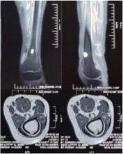
          19-year-old female presented with persistent pain on her left thigh for 4 years. Aside from tenderness on her left thigh, her physical examination was unremarkable. Plain radiographic of left femur showed a small geographic osteolytic cortical lesion with sclerotic rim in the distal region. Further evaluation with MRI showed eccentric lytic lesion with an isointense signal on T1-weighted images and a hyperintense signals on T2-weighted images. Patient then temporarily diagnosed with osteoblastoma. Because there were no signs of malignancy, the patient underwent a curettage of the mass followed by synthetic bone graft application. Histopathological findings were consistent for schwannoma. Further immunohistochemical examination showed positive S100 staining, confirming the final diagnosis of intraosseous schwannoma. There were no signs of early complication on 3 months post-operation. The patient was further scheduled for follow up on 6 months and then routinely every year post-operation to evaluate any signs of complication or recurrence.

          Clinical discussion

          It is difficult to make an accurate initial diagnosis of intraosseous schwannomas. Because the clinical presentation was most likely not specific as such in this case and there many other tumors of the bone with similar radiographic finding which are more common. Curettage of the mass followed by synthetic bone graft application was performed as there were no sign of malignancy making more invasive option deemed to cause more harm than good to the patient.

          Conclusion

          The possibility of intraosseous schwannoma should have been taken under consideration in the differential diagnosis of osteolytic lesion with pain in long bone.

          Highlights

          • Clinical presentation of intraosseous schwannoma can be non-specific

          • Radiological findings of intraosseous schwannoma can mimic more common condition of the bone such as osteoblastoma, non-ossifying fibroma, and simple bone cyst

          • Histopathological and immunohistochemical analysis is important to established the final diagnosis

          • Curettage of the mass followed by synthetic bone graft application are adequate for intraosseous schwannoma as it is a benign condition with almost negligible potential to become malignant.

          Related collections

          Most cited references10

          • Record: found
          • Abstract: found
          • Article: not found

          The SCARE 2020 Guideline: Updating Consensus Surgical CAse REport (SCARE) Guidelines

          The SCARE Guidelines were first published in 2016 and were last updated in 2018. They provide a structure for reporting surgical case reports and are used and endorsed by authors, journal editors and reviewers, in order to increase robustness and transparency in reporting surgical cases. They must be kept up to date in order to drive forwards reporting quality. As such, we have updated these guidelines via a DELPHI consensus exercise.
            Bookmark
            • Record: found
            • Abstract: found
            • Article: found
            Is Open Access

            Airborne transmission of COVID-19 and the role of face mask to prevent it: a systematic review and meta-analysis

            Background and aims Severe acute respiratory syndrome coronavirus 2 (SARS-CoV-2), belonging to the Coronaviridae family, is agent of 2019 novel coronavirus disease (COVID-19). COVID-19 emerged in Wuhan, Hubei province of China, in early December 2019 and is now considered a pandemic. This study aimed to investigate the airborne transmission of COVID-19 and the role of face mask to prevent it. Methods A systematic search for English-language literature was done via PUBMED/Medline and Google Scholar up to October 2020. There was two search strategy; for airborne transmission and the role of face mask for prevention of SARS-CoV-2 infection. Based on a fixed and random effects model, the RR and 95% CI were used to evaluate the combined risk. This meta-analysis followed Preferred Reporting Items for Systematic Reviews and Meta-analysis (PRISMA) Guidelines. Results After eligibility assessment, four articles with a total of 7688 participants were included in this meta-analysis. The result of this meta-analysis has shown significant reduction in infection with face mask use; the pooled RR (95%CI) was 0.12 [0.06, 0.27] (P < 0.001). Conclusion In conclusion, this meta-analysis suggests that there is association between face mask use and reduction of COVID-19. However, COVID-19 spreads primarily with contact routes and respiratory droplets, but its transmissibility has many mysteries yet and there is controversy about airborne transmission of COVID-19.
              Bookmark
              • Record: found
              • Abstract: found
              • Article: not found

              Association between intraosseous schwannoma occurrence and the position of the intraosseous nutrient vessel: A case report.

              Intraosseous schwannoma is a rare benign bone tumor that originates from Schwann cells of the nerve sheath. The majority of intraosseous schwannomas arise in the mandible and sacrum, but an intraosseous schwannoma involving the ulna is described in the present case report. Radiologically, the current case presented as a well-defined lytic lesion, with a pathological fracture and no intralesional calcification, in the proximal metaphysis of the left ulna. Using magnetic resonance imaging, an intraosseous mass spreading out from the cortical defect was observed. The lesion appeared isointense to skeletal muscle on T1-weighted images, and hyperintense or heterogeneous on T2-weighted images. The differential diagnosis comprised benign bone tumors, including bone cyst, aneurysmal bone cyst, giant cell tumor and fibrous dysplasia. Based on the results of a needle biopsy, a schwannoma involving the ulnar bone was diagnosed, and tumor marginal resection followed by artificial bone grafting and fixation was performed. A total of one year subsequent to surgery, the patient exhibits no symptoms, and there is no evidence of disease recurrence.
                Bookmark

                Author and article information

                Contributors
                Journal
                Int J Surg Case Rep
                Int J Surg Case Rep
                International Journal of Surgery Case Reports
                Elsevier
                2210-2612
                13 September 2022
                October 2022
                13 September 2022
                : 99
                : 107643
                Affiliations
                [a ]Department of Orthopaedic and Traumatology, Fatmawati General Hospital, Jakarta, Indonesia
                [b ]Fatmawati General Hospital, Jakarta, Indonesia
                [c ]Department of Anatomical Pathology, Fatmawati General Hospital, Jakarta, Indonesia
                Author notes
                [* ]Corresponding author. rama.premiarto@ 123456gmail.com
                Article
                S2210-2612(22)00889-6 107643
                10.1016/j.ijscr.2022.107643
                9568785
                36174461
                bdcd2fb1-2fe6-4641-8a53-80adc237b987
                © 2022 The Authors

                This is an open access article under the CC BY-NC-ND license (http://creativecommons.org/licenses/by-nc-nd/4.0/).

                History
                : 3 July 2022
                : 15 August 2022
                : 9 September 2022
                Categories
                Case Report

                case report,femur,intraosseous,primary,schwannoma,tumor
                case report, femur, intraosseous, primary, schwannoma, tumor

                Comments

                Comment on this article