538
views
0
recommends
+1 Recommend
0 collections
    0
    shares
      • Record: found
      • Abstract: found
      • Article: not found

      Ulnar-sided wrist pain. II. Clinical imaging and treatment

      review-article

      Read this article at

      ScienceOpenPublisherPMC
          There is no author summary for this article yet. Authors can add summaries to their articles on ScienceOpen to make them more accessible to a non-specialist audience.

          Abstract

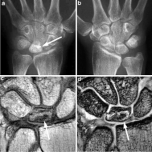
          Pain at the ulnar aspect of the wrist is a diagnostic challenge for hand surgeons and radiologists due to the small and complex anatomical structures involved. In this article, imaging modalities including radiography, arthrography, ultrasound (US), computed tomography (CT), CT arthrography, magnetic resonance (MR) imaging, and MR arthrography are compared with regard to differential diagnosis. Clinical imaging findings are reviewed for a more comprehensive understanding of this disorder. Treatments for the common diseases that cause the ulnar-sided wrist pain including extensor carpi ulnaris (ECU) tendonitis, flexor carpi ulnaris (FCU) tendonitis, pisotriquetral arthritis, triangular fibrocartilage complex (TFCC) lesions, ulnar impaction, lunotriquetral (LT) instability, and distal radioulnar joint (DRUJ) instability are reviewed.

          Related collections

          Most cited references126

          • Record: found
          • Abstract: found
          • Article: not found

          Triangular fibrocartilage complex lesions: a classification.

          Based on anatomic and biomechanical studies and review of our clinical experience of the past 10 years, a classification of injuries to the triangular fibrocartilage complex is presented. This classification is based on the clinical examination, routine x-ray films, wrist arthrograms, wrist arthroscopy, and wrist arthrotomy. The classification recognizes both traumatic and degenerative lesions. Traumatic lesions are classified according to their location. Degenerative lesions are classified by the location and severity of degenerative changes of the triangular fibrocartilage complex, ulnar head, ulnocarpal bones and lunotriquetral ligament.
            Bookmark
            • Record: found
            • Abstract: found
            • Article: not found

            Wrist fractures: what the clinician wants to know.

            With the recent improvements in diagnosis and treatment of distal radius and carpal injuries, the hand surgeons' expectations of relevant radiologic interpretation of imaging studies are heightened. Conventional radiographic examinations, as well as more sophisticated and invasive studies, have important roles in the evaluation of wrist fractures and dislocations. On the basis of physical examination results and the mechanism of injury, the onus is on the examining surgeon to pinpoint potential sites of bone or ligament disruption. After this evaluation, appropriate imaging studies appropriately performed and interpreted will help direct treatment and improve outcome with greater clarity and certainty.
              Bookmark
              • Record: found
              • Abstract: found
              • Article: not found

              Special focus session. MR arthrography.

              Direct magnetic resonance (MR) arthrography with injection of saline solution or diluted gadolinium can be useful for evaluating certain pathologic conditions in the joints. It is most helpful for outlining labral-ligamentous abnormalities in the shoulder and distinguishing partial-thickness from full-thickness tears in the rotator cuff, demonstrating labral tears in the hip, showing partial- and full-thickness tears of the collateral ligament of the elbow and delineating bands in the elbow, identifying residual or recurrent tears in the knee following meniscectomy, increasing the certainty of perforations of the ligaments and triangular fibrocartilage in the wrist, correctly identifying ligament tears in the ankle and increasing the sensitivity for ankle impingement syndromes, assessing the stability of osteochondral lesions in the articular surface of joints, and delineating loose bodies in joints. Indirect MR arthrography with intravenous administration of diluted gadolinium may be performed when direct arthrography is inconvenient or not logistically feasible. Although indirect MR arthrography has some disadvantages vis-à-vis direct MR arthrography, it does not require fluoroscopic guidance or joint injection and it is superior to conventional MR imaging in delineating structures when there is minimal joint fluid. In addition, vascularized or inflamed tissue will enhance with this method. Indirect MR arthrography can be used to rule in or diagnose abnormalities and to exclude abnormalities.
                Bookmark

                Author and article information

                Contributors
                +1-714-4568849 , +1-714-4567430 , hiroshi@uci.edu
                Journal
                Skeletal Radiol
                Skeletal Radiology
                Springer-Verlag (Berlin/Heidelberg )
                0364-2348
                1432-2161
                10 December 2009
                10 December 2009
                September 2010
                : 39
                : 9
                : 837-857
                Affiliations
                [1 ]Department of Radiology, Brigham and Women’s Hospital, Boston, MA USA
                [2 ]Department of Orthopaedic Surgery, Brigham and Women’s Hospital, Boston, MA USA
                [3 ]Department of Radiological Sciences, University of California-Irvine, Irvine, CA USA
                [4 ]Department of Radiological Sciences, UC Irvine Medical Center, 101 City Drive South, Route 140, Orange, CA 92868 USA
                Article
                842
                10.1007/s00256-009-0842-3
                2904904
                20012039
                fb2c7641-f904-4dbc-82ca-674cfc8839c3
                © The Author(s) 2009
                History
                : 29 June 2009
                : 14 November 2009
                : 17 November 2009
                Categories
                Review Article
                Custom metadata
                © ISS 2010

                Radiology & Imaging
                ulnar side,wrist pain,imaging,treatment
                Radiology & Imaging
                ulnar side, wrist pain, imaging, treatment

                Comments

                Comment on this article