73
views
0
recommends
+1 Recommend
0 collections
    2
    shares
      • Record: found
      • Abstract: found
      • Article: found
      Is Open Access

      A Novel Role for SIRT-1 in L-Arginine Protection against STZ Induced Myocardial Fibrosis in Rats

      research-article
      1 , 1 , 2 , *
      PLoS ONE
      Public Library of Science

      Read this article at

          There is no author summary for this article yet. Authors can add summaries to their articles on ScienceOpen to make them more accessible to a non-specialist audience.

          Abstract

          Background

          L-arginine (L-ARG) effectively protects against diabetic impediments. In addition, silent information regulator (SIRT-1) activators are emerging as a new clinical concept in treating diabetic complications. Accordingly, this study aimed at delineating a role for SIRT-1 in mediating L-ARG protection against streptozotocin (STZ) induced myocardial fibrosis.

          Methods

          Male Wistar rats were allocated into five groups; (i) normal control rats received 0.1 M sodium citrate buffer (pH 4.5); (ii) STZ at the dose of 60 mg/kg dissolved in 0.1 M sodium citrate buffer (pH 4.5); (iii) STZ + sirtinol (Stnl; specific inhibitor of SIRT-1; 2 mg/Kg, i.p.); (iv) STZ + L-ARG given in drinking water (2.25%) or (v) STZ + L-ARG + Stnl.

          Results

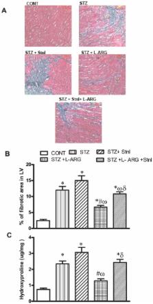
          L-ARG increased myocardial SIRT-1 expression as well as its protein content. The former finding was paralleled by L-ARG induced reduction in myocardial fibrotic area compared to STZ animals evidenced histopathologically. The reduction in the fibrotic area was accompanied by a decline in fibrotic markers as evident by a decrease in expression of collagen-1 along with reductions in myocardial TGF-β, fibronectin, CTGF and BNP expression together with a decrease in TGF-β and hydroxyproline contents. Moreover, L-ARG increased MMP-2 expression in addition to its protein content while decreasing expression of PAI-1. Finally, L-ARG protected against myocardial cellular death by reduction in NFκ-B mRNA as well as TNF-α level in association with decline in Casp-3 and FAS expressions andCasp-3protein content in addition to reduction of FAS positive cells. However, co-administration of L-ARG and Stnl diminished the protective effect of L-ARG against STZ induced myocardial fibrosis.

          Conclusion

          Collectively, these findings associate a role for SIRT-1 in L-ARG defense against diabetic cardiac fibrosis via equilibrating the balance between profibrotic and antifibrotic mediators.

          Related collections

          Most cited references60

          • Record: found
          • Abstract: found
          • Article: not found

          Potential therapeutic targets for cardiac fibrosis: TGFbeta, angiotensin, endothelin, CCN2, and PDGF, partners in fibroblast activation.

          Fibrosis is one of the largest groups of diseases for which there is no therapy but is believed to occur because of a persistent tissue repair program. During connective tissue repair, "activated" fibroblasts migrate into the wound area, where they synthesize and remodel newly created extracellular matrix. The specialized type of fibroblast responsible for this action is the alpha-smooth muscle actin (alpha-SMA)-expressing myofibroblast. Abnormal persistence of the myofibroblast is a hallmark of fibrotic diseases. Proteins such as transforming growth factor (TGF)beta, endothelin-1, angiotensin II (Ang II), connective tissue growth factor (CCN2/CTGF), and platelet-derived growth factor (PDGF) appear to act in a network that contributes to myofibroblast differentiation and persistence. Drugs targeting these proteins are currently under consideration as antifibrotic treatments. This review summarizes recent observations concerning the contribution of TGFbeta, endothelin-1, Ang II, CCN2, and PDGF and to fibroblast activation in tissue repair and fibrosis and the potential utility of agents blocking these proteins in affecting the outcome of cardiac fibrosis.
            Bookmark
            • Record: found
            • Abstract: found
            • Article: not found

            Redox regulation of SIRT1 in inflammation and cellular senescence.

            Sirtuin 1 (SIRT1) regulates inflammation, aging (life span and health span), calorie restriction/energetics, mitochondrial biogenesis, stress resistance, cellular senescence, endothelial functions, apoptosis/autophagy, and circadian rhythms through deacetylation of transcription factors and histones. SIRT1 level and activity are decreased in chronic inflammatory conditions and aging, in which oxidative stress occurs. SIRT1 is regulated by a NAD(+)-dependent DNA repair enzyme, poly(ADP-ribose) polymerase-1 (PARP1), and subsequent NAD(+) depletion by oxidative stress may have consequent effects on inflammatory and stress responses as well as cellular senescence. SIRT1 has been shown to undergo covalent oxidative modifications by cigarette smoke-derived oxidants/aldehydes, leading to posttranslational modifications, inactivation, and protein degradation. Furthermore, oxidant/carbonyl stress-mediated reduction of SIRT1 leads to the loss of its control on acetylation of target proteins including p53, RelA/p65, and FOXO3, thereby enhancing the inflammatory, prosenescent, and apoptotic responses, as well as endothelial dysfunction. In this review, the mechanisms of cigarette smoke/oxidant-mediated redox posttranslational modifications of SIRT1 and its roles in PARP1 and NF-κB activation, and FOXO3 and eNOS regulation, as well as chromatin remodeling/histone modifications during inflammaging, are discussed. Furthermore, we have also discussed various novel ways to activate SIRT1 either directly or indirectly, which may have therapeutic potential in attenuating inflammation and premature senescence involved in chronic lung diseases. Copyright © 2013 Elsevier Inc. All rights reserved.
              Bookmark
              • Record: found
              • Abstract: found
              • Article: not found

              TGF-beta and fibrosis in different organs - molecular pathway imprints.

              The action of transforming-growth-factor (TGF)-beta following inflammatory responses is characterized by increased production of extracellular matrix (ECM) components, as well as mesenchymal cell proliferation, migration, and accumulation. Thus, TGF-beta is important for the induction of fibrosis often associated with chronic phases of inflammatory diseases. This common feature of TGF-related pathologies is observed in many different organs. Therefore, in addition to the description of the common TGF-beta-pathway, this review focuses on TGF-beta-related pathogenetic effects in different pathologies/organs, i. e., arthritis, diabetic nephropathy, colitis/Crohn's disease, radiation-induced fibrosis, and myocarditis (including their similarities and dissimilarities). However, TGF-beta exhibits both exacerbating and ameliorating features, depending on the phase of disease and the site of action. Due to its central role in severe fibrotic diseases, TGF-beta nevertheless remains an attractive therapeutic target, if targeted locally and during the fibrotic phase of disease.
                Bookmark

                Author and article information

                Contributors
                Role: Editor
                Journal
                PLoS One
                PLoS ONE
                plos
                plosone
                PLoS ONE
                Public Library of Science (San Francisco, USA )
                1932-6203
                2014
                12 December 2014
                : 9
                : 12
                : e114560
                Affiliations
                [1 ]Department of Biochemistry, Faculty of Pharmacy, Cairo University, Cairo, Egypt
                [2 ]Department of Pharmacology and Toxicology, Faculty of Pharmacy, Cairo University, Cairo, Egypt
                University of Central Florida, United States of America
                Author notes

                Competing Interests: The authors have declared that no competing interests exist.

                Conceived and designed the experiments: SMR SAE NNN. Performed the experiments: SMR SAE NNN. Analyzed the data: SMR SAE NNN. Contributed reagents/materials/analysis tools: SMR SAE NNN. Wrote the paper: SMR SAE NNN.

                Article
                PONE-D-14-33592
                10.1371/journal.pone.0114560
                4264750
                25501750
                888ef1cb-d052-4af5-b257-157bd68a9d9e
                Copyright @ 2014

                This is an open-access article distributed under the terms of the Creative Commons Attribution License, which permits unrestricted use, distribution, and reproduction in any medium, provided the original author and source are credited.

                History
                : 26 July 2014
                : 11 November 2014
                Page count
                Pages: 19
                Funding
                These authors have no support or funding to report.
                Categories
                Research Article
                Biology and Life Sciences
                Organisms
                Animals
                Vertebrates
                Mammals
                Rodents
                Physiology
                Cardiovascular Physiology
                Custom metadata
                The authors confirm that all data underlying the findings are fully available without restriction. All relevant data are within the paper.

                Uncategorized
                Uncategorized

                Comments

                Comment on this article