- Record: found
- Abstract: found
- Article: not found
Asymmetric distribution and spatial switching of dynein activity generates ciliary motility
Read this article at

Abstract
Motile cilia and flagella are essential, highly conserved organelles, and their motility is driven by the coordinated activities of multiple dynein isoforms. The prevailing “switch-point” hypothesis posits that dyneins are asymmetrically activated to drive flagellar bending. To test this model, we applied cryo–electron tomography to visualize activity states of individual dyneins relative to their locations along beating flagella of sea urchin sperm cells. As predicted, bending was generated by the asymmetric distribution of dynein activity on opposite sides of the flagellum. However, contrary to predictions, most dyneins were in their active state, and the smaller population of conformationally inactive dyneins switched flagellar sides relative to the bending direction. Thus, our data suggest a “switch-inhibition” mechanism in which force imbalance is generated by inhibiting, rather than activating, dyneins on alternating sides of the flagellum.
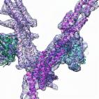
Graphical Abstract
INTRODUCTION:
Motile cilia and flagella are highly conserved, hairlike appendages of eukaryotic cells that propel the movement of cells or fluids. They play important roles in the normal development and health of many species, including humans. Flagellar beating is driven by the coordinated activities of multiple dynein isoforms that must be spatially and temporally regulated. Although the prevailing “switch-point” hypothesis posits that flagellar motility results from periodic switching of spatially restricted, asymmetrical activation of dyneins, no direct evidence has been reported, and how the thousands of dyneins inside a flagellum work together to generate flagellar motility remains elusive.
RATIONALE:
Here we rapidly froze swimming sea urchin sperm cells and used cryo–electron tomography (cryo-ET) to image their beating flagella. Subtomogram averaging and classification analyses allowed us to identify and visualize the different activity states of individual dyneins and their regulators in situ. These conformational states were then mapped to their locations along the sinusoidal wave of the beating flagellum, for example, in relation to principal bend, reverse bend, or straight regions between bends. The results allowed us to elucidate the distinct roles played by various dyneins and to propose a model for the mechanism that underlies ciliary and flagellar motility.
RESULTS:
The native three-dimensional structures of flagellar complexes were determined in situ with resolutions sufficient for identifying different activity states. Dyneins of immotile control flagella were found to be in post–power stroke conformations (unprimed, inactive states). By contrast, in beating flagella, most dyneins were in pre–power stroke conformations (primed, active states), with only a few dyneins in intermediate conformations. Moreover, for all outer dyneins, the intermediate and inactive conformations were only found in bent regions and were clustered on one side of the flagellum in a bend direction–dependent manner. For inner dyneins, certain isoforms (dyneins I1, a, d, and g) showed similar bend direction–dependent distribution patterns in bent regions of flagella, whereas the distribution patterns of other isoforms (dyneins b, c, and e) lacked obvious correlations with bending direction.
Our results revealed three key tenets that are important for generating flagellar motility: (i) The asymmetric distribution of dynein activity on opposite sides of the flagellum results in unidirectional bending, and (ii) the switching of dynein conformations between opposite sides causes the undulating waveform of beating flagella, both of which directly confirmed the switching aspect of the previously proposed switch-point hypothesis. (iii) In contrast to predictions, however, the findings also suggested the paradigm-shifting model that dyneins are active by default and that the asymmetry of dynein activity is driven by spatially restricted inhibition rather than activation of dyneins on alternating sides of the flagellum. This “switch-inhibition” mechanism was further supported by our analyses of a regulation-deficient Chlamydomonas mutant, which revealed that dyneins consumed adenosine triphosphate (ATP) and adopted pre–power stroke conformations, even though flagella were paralyzed.
CONCLUSION:
Our comprehensive structural analysis combined with biochemical investigations provides an enhanced understanding of the distinct roles played by various dyneins and regulatory complexes in the motility of cilia and flagella and suggests critical modifications to previous hypotheses regarding robust molecular mechanisms underlying flagellar motility. Our study demonstrates that comparative cellular cryo-ET studies provide the conceptual framework and experimental tools to better understand molecular mechanisms and cellular functions.

Asymmetric dynein activity underlies beating of cilia and flagella. Cryo-ET was used to image the active flagellum of swimming sea urchin sperm cells. Different activity states of the motility-driving dynein motors were identified. Magnified views show active (right) and inactive intermediate states (left). The distribution patterns of dynein conformations along the undulating waveform suggest a switch-inhibition mechanism for ciliary and flagellar motility.
Related collections
Most cited references40
- Record: found
- Abstract: found
- Article: not found
Functions and mechanics of dynein motor proteins.
- Record: found
- Abstract: found
- Article: found
Cryo-EM Reveals How Human Cytoplasmic Dynein Is Auto-inhibited and Activated
- Record: found
- Abstract: found
- Article: not found