20
views
0
recommends
+1 Recommend
0 collections
    0
    shares
      • Record: found
      • Abstract: found
      • Article: not found

      Comparison of Carboplatin and Doxorubicin‐Based Chemotherapy Protocols in 470 Dogs after Amputation for Treatment of Appendicular Osteosarcoma

      research-article

      Read this article at

      ScienceOpenPublisherPMC
          There is no author summary for this article yet. Authors can add summaries to their articles on ScienceOpen to make them more accessible to a non-specialist audience.

          Abstract

          Background

          Many chemotherapy protocols have been reported for treatment of canine appendicular osteosarcoma ( OSA), but outcome comparisons in a single population are lacking.

          Objective

          To evaluate the effects of protocol and dose intensity ( DI) on treatment outcomes for carboplatin and doxorubicin‐based chemotherapy protocols.

          Animals

          Four hundred and seventy dogs with appendicular OSA.

          Methods

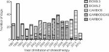
          A retrospective cohort study was performed comprising consecutive dogs treated (1997–2012) with amputation followed by 1 of 5 chemotherapy protocols: carboplatin 300 mg/m 2 IV q21d for 4 or 6 cycles ( CARBO6), doxorubicin 30 mg/m 2 IV q14d or q21d for 5 cycles, and alternating carboplatin 300 mg/m 2 IV and doxorubicin 30 mg/m 2 IV q21d for 3 cycles. Adverse events ( AE) and DI were evaluated. Kaplan–Meier survival curves and Cox proportional hazards regression were used to compare disease‐free interval ( DFI) and survival time ( ST) among protocols.

          Results

          The overall median DFI and ST were 291 days and 284 days, respectively. A lower proportion of dogs prescribed CARBO6 experienced AEs compared to other protocols (48.4% versus 60.8–75.8%; P = .001). DI was not associated with development of metastases or death. After adjustment for baseline characteristics and prognostic factors, none of the protocols provided a significant reduction in risk of development of metastases or death.

          Conclusions and Clinical Importance

          Although choice of protocol did not result in significant differences in DFI or ST, the CARBO6 protocol resulted in a lower proportion of dogs experiencing AEs, which could be advantageous in maintaining high quality of life during treatment. DI was not a prognostic indicator in this study.

          Related collections

          Most cited references29

          • Record: found
          • Abstract: found
          • Article: not found

          Improved survival associated with postoperative wound infection in dogs treated with limb-salvage surgery for osteosarcoma.

          Limb-salvage surgery and adjuvant chemotherapy are performed as a treatment of appendicular osteosarcoma in dogs. Approximately 50% of dogs that undergo limb-salvage surgery develop postoperative surgical wound infections. Postoperative surgical infections may affect survival in cancer patients. The purposes of this study were to examine the effect of surgical wound infection on survival, local recurrence, and metastasis in relation to other prognostic factors for dogs with spontaneous osteosarcoma treated with limb-salvage surgery. Forty-seven client-owned dogs with osteosarcoma of the distal radius were treated with limb-salvage surgery and adjuvant chemotherapy--either carboplatin or carboplatin and doxorubicin. Hazard ratios were estimated by using the Cox proportional hazard model, and survival functions were estimated by using the Kaplan-Meier product-limit life-table method. Of the 47 dogs in this study, 32 (68%) developed a postoperative wound infection. Infection, dog weight, and extent of the primary tumor (percentage of length) significantly affected survival, and infection and percentage of length significantly affected time to metastasis. None of the variables considered in this study affected local recurrence. Dogs that were diagnosed with an infection were less likely to die (hazard ratio, .446), and dogs with greater body weight and greater percentage length involvement were more likely to die (hazard ratios of 3.37 and 3.66, respectively). In dogs with osteosarcoma treated with limb-salvage surgery, infection has a positive influence on survival, as does a smaller initial length of radius involved and lower body weight.
            Bookmark
            • Record: found
            • Abstract: found
            • Article: found
            Is Open Access

            Prognostic factors in canine appendicular osteosarcoma – a meta-analysis

            Background Appendicular osteosarcoma is the most common malignant primary canine bone tumor. When treated by amputation or tumor removal alone, median survival times (MST) do not exceed 5 months, with the majority of dogs suffering from metastatic disease. This period can be extended with adequate local intervention and adjuvant chemotherapy, which has become common practice. Several prognostic factors have been reported in many different studies, e.g. age, breed, weight, sex, neuter status, location of tumor, serum alkaline phosphatase (SALP), bone alkaline phosphatase (BALP), infection, percentage of bone length affected, histological grade or histological subtype of tumor. Most of these factors are, however, only reported as confounding factors in larger studies. Insight in truly significant prognostic factors at time of diagnosis may contribute to tailoring adjuvant therapy for individual dogs suffering from osteosarcoma. The objective of this study was to systematically review the prognostic factors that are described for canine appendicular osteosarcoma and validate their scientific importance. Results A literature review was performed on selected studies and eligible data were extracted. Meta-analyses were done for two of the three selected possible prognostic factors (SALP and location), looking at both survival time (ST) and disease free interval (DFI). The third factor (age) was studied in a qualitative manner. Both elevated SALP level and the (proximal) humerus as location of the primary tumor are significant negative prognostic factors for both ST and DFI in dogs with appendicular osteosarcoma. Increasing age was associated with shorter ST and DFI, however, was not statistically significant because information of this factor was available in only a limited number of papers. Conclusions Elevated SALP and proximal humeral location are significant negative prognosticators for canine osteosarcoma.
              Bookmark
              • Record: found
              • Abstract: found
              • Article: not found

              Use of single-agent carboplatin as adjuvant or neoadjuvant therapy in conjunction with amputation for appendicular osteosarcoma in dogs.

              Survival following amputation and administration of single-agent carboplatin for treatment of appendicular osteosarcoma (OSA) in dogs was retrospectively examined. Records of 155 dogs with appendicular OSA treated with amputation and single-agent carboplatin were included from 14 centers. Any carboplatin dosage, number of doses, and protocol schedule were eligible for inclusion. The median disease-free interval (DFI) was 256 days. The median overall survival time was 307 days. Similar prognostic survival factors were identified in this study as reported in prior studies of canine appendicular OSA. Median DFI and survival were comparable to those reported in the original Bergman et al publication. Carboplatin treatment improves the survival probability in dogs with appendicular OSA compared to amputation alone and remains an acceptable alternative to adjuvant treatment with cisplatin.
                Bookmark

                Author and article information

                Journal
                J Vet Intern Med
                J. Vet. Intern. Med
                10.1111/(ISSN)1939-1676
                JVIM
                Journal of Veterinary Internal Medicine
                John Wiley and Sons Inc. (Hoboken )
                0891-6640
                1939-1676
                10 February 2014
                Mar-Apr 2014
                : 28
                : 2 ( doiID: 10.1111/jvim.2014.28.issue-2 )
                : 554-563
                Affiliations
                [ 1 ] Flint Animal Cancer Center Department of Clinical Sciences College of Veterinary Medicine and Biomedical SciencesColorado State University Fort Collins CO
                Author notes
                [*] [* ]Corresponding author: L.E. Selmic, BVetMed (Hons) DACVS‐SA, ACVS Founding Fellow of Surgical Oncology, The Flint Animal Cancer Center, Department of Clinical Sciences, College of Veterinary Medicine and Biomedical Sciences, Colorado State University, 300 W. Drake Road, Fort Collins, CO 80523; email: laura.selmic@ 123456gmail.com
                Article
                JVIM12313
                10.1111/jvim.12313
                4857984
                24512451
                1f576370-3a03-4417-b2ab-5d3d63092da4
                Copyright © 2014 by the American College of Veterinary Internal Medicine
                History
                : 13 August 2013
                : 26 November 2013
                : 26 December 2013
                Page count
                Pages: 10
                Categories
                Original Article
                Standard Articles
                Custom metadata
                2.0
                jvim12313
                March/April 2014
                Converter:WILEY_ML3GV2_TO_NLMPMC version:4.8.8 mode:remove_FC converted:25.04.2016

                Veterinary medicine
                canine,limb amputation,oncology,primary bone tumor
                Veterinary medicine
                canine, limb amputation, oncology, primary bone tumor

                Comments

                Comment on this article