21
views
0
recommends
+1 Recommend
0 collections
    0
    shares
      • Record: found
      • Abstract: found
      • Article: found
      Is Open Access

      Astrocyte Unfolded Protein Response Induces a Specific Reactivity State that Causes Non-Cell-Autonomous Neuronal Degeneration

      research-article

      Read this article at

          There is no author summary for this article yet. Authors can add summaries to their articles on ScienceOpen to make them more accessible to a non-specialist audience.

          Summary

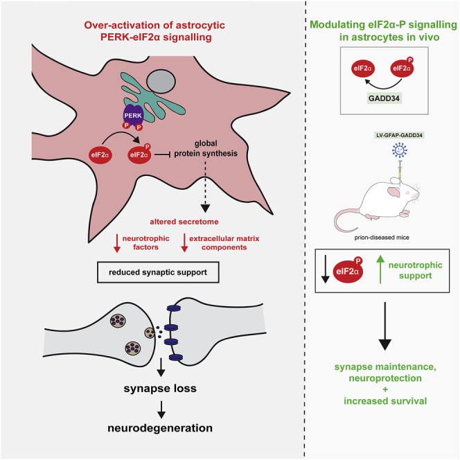
          Recent interest in astrocyte activation states has raised the fundamental question of how these cells, normally essential for synapse and neuronal maintenance, become pathogenic. Here, we show that activation of the unfolded protein response (UPR), specifically phosphorylated protein kinase R-like endoplasmic reticulum (ER) kinase (PERK-P) signaling—a pathway that is widely dysregulated in neurodegenerative diseases—generates a distinct reactivity state in astrocytes that alters the astrocytic secretome, leading to loss of synaptogenic function in vitro. Further, we establish that the same PERK-P-dependent astrocyte reactivity state is harmful to neurons in vivo in mice with prion neurodegeneration. Critically, targeting this signaling exclusively in astrocytes during prion disease is alone sufficient to prevent neuronal loss and significantly prolongs survival. Thus, the astrocyte reactivity state resulting from UPR over-activation is a distinct pathogenic mechanism that can by itself be effectively targeted for neuroprotection.

          Graphical Abstract

          Highlights

          • PERK-eIF2α signaling in astrocytes generates a distinct “UPR”-reactivity state

          • UPR-reactive astrocytes have an altered secretome, with reduced synaptogenic factors

          • UPR-reactive astrocytes fail to support synaptogenesis in vitro

          • Targeting the astrocytic UPR prevents synapse and neuronal loss in prion-diseased mice

          Abstract

          Dysregulation of UPR signaling in neurons is a key mediator of neurodegeneration. Smith et al. show that UPR dysregulation in astrocytes impairs their ability to support synapses; modulating astrocytic UPR signaling prevents neuronal loss and increases survival in prion-diseased mice.

          Related collections

          Most cited references29

          • Record: found
          • Abstract: found
          • Article: not found

          Thrombospondins are astrocyte-secreted proteins that promote CNS synaptogenesis.

          The establishment of neural circuitry requires vast numbers of synapses to be generated during a specific window of brain development, but it is not known why the developing mammalian brain has a much greater capacity to generate new synapses than the adult brain. Here we report that immature but not mature astrocytes express thrombospondins (TSPs)-1 and -2 and that these TSPs promote CNS synaptogenesis in vitro and in vivo. TSPs induce ultrastructurally normal synapses that are presynaptically active but postsynaptically silent and work in concert with other, as yet unidentified, astrocyte-derived signals to produce functional synapses. These studies identify TSPs as CNS synaptogenic proteins, provide evidence that astrocytes are important contributors to synaptogenesis within the developing CNS, and suggest that TSP-1 and -2 act as a permissive switch that times CNS synaptogenesis by enabling neuronal molecules to assemble into synapses within a specific window of CNS development.
            Bookmark
            • Record: found
            • Abstract: found
            • Article: not found

            Sustained translational repression by eIF2α-P mediates prion neurodegeneration.

            The mechanisms leading to neuronal death in neurodegenerative disease are poorly understood. Many of these disorders, including Alzheimer's, Parkinson's and prion diseases, are associated with the accumulation of misfolded disease-specific proteins. The unfolded protein response is a protective cellular mechanism triggered by rising levels of misfolded proteins. One arm of this pathway results in the transient shutdown of protein translation, through phosphorylation of the α-subunit of eukaryotic translation initiation factor, eIF2. Activation of the unfolded protein response and/or increased eIF2α-P levels are seen in patients with Alzheimer's, Parkinson's and prion diseases, but how this links to neurodegeneration is unknown. Here we show that accumulation of prion protein during prion replication causes persistent translational repression of global protein synthesis by eIF2α-P, associated with synaptic failure and neuronal loss in prion-diseased mice. Further, we show that promoting translational recovery in hippocampi of prion-infected mice is neuroprotective. Overexpression of GADD34, a specific eIF2α-P phosphatase, as well as reduction of levels of prion protein by lentivirally mediated RNA interference, reduced eIF2α-P levels. As a result, both approaches restored vital translation rates during prion disease, rescuing synaptic deficits and neuronal loss, thereby significantly increasing survival. In contrast, salubrinal, an inhibitor of eIF2α-P dephosphorylation, increased eIF2α-P levels, exacerbating neurotoxicity and significantly reducing survival in prion-diseased mice. Given the prevalence of protein misfolding and activation of the unfolded protein response in several neurodegenerative diseases, our results suggest that manipulation of common pathways such as translational control, rather than disease-specific approaches, may lead to new therapies preventing synaptic failure and neuronal loss across the spectrum of these disorders.
              Bookmark
              • Record: found
              • Abstract: found
              • Article: not found

              Astrocyte glypicans 4 and 6 promote formation of excitatory synapses via GluA1 AMPA receptors

              In the developing central nervous system (CNS), the control of synapse number and function is critical to the formation of neural circuits. We previously demonstrated that astrocyte-secreted factors powerfully induce the formation of functional excitatory synapses between CNS neurons 1 . Astrocyte-secreted thrombospondins induce structural synapses, however these synapses are post-synaptically silent 2 . Here we use biochemical fractionation of astrocyte conditioned media (ACM) to identify glypican 4 (Gpc4) and 6 (Gpc6) as astrocyte-secreted signals sufficient to induce functional synapses between purified retinal ganglion cell (RGC) neurons, and show that depletion of these molecules from ACM significantly reduces its ability to induce postsynaptic activity. Application of Gpc4 to purified neurons is sufficient to increase the frequency and amplitude of glutamatergic synaptic events. This is achieved by increasing the surface level and clustering, but not overall cellular protein level, of the GluA1 subunit of the AMPA glutamate receptor (AMPAR). Gpc4&6 are expressed by astrocytes in vivo in the developing CNS, with Gpc4 expression enriched in the hippocampus and Gpc6 in the cerebellum. Finally, we demonstrate that Gpc4-deficient mice have defective synapse formation, with decreased amplitude of excitatory synaptic currents in the developing hippocampus and reduced recruitment of AMPARs to synapses. These data identify glypicans as a family of novel astrocyte-derived molecules that are necessary and sufficient to promote glutamate receptor clustering and receptivity and induce the formation of post-synaptically functioning CNS synapses.
                Bookmark

                Author and article information

                Contributors
                Journal
                Neuron
                Neuron
                Neuron
                Cell Press
                0896-6273
                1097-4199
                04 March 2020
                04 March 2020
                : 105
                : 5
                : 855-866.e5
                Affiliations
                [1 ]Department of Clinical Neurosciences and UK Dementia Research Institute at the University of Cambridge, Island Research Building, Cambridge Biomedical Campus, Cambridge, UK
                [2 ]Wellcome-MRC Cambridge Stem Cell Institute, Jeffrey Cheah Biomedical Centre, Cambridge Biomedical Campus, Cambridge, UK
                [3 ]MRC Toxicology Unit, Hodgkin Building, Leicester, UK
                Author notes
                []Corresponding author gm522@ 123456cam.ac.uk
                [4]

                Lead Contact

                Article
                S0896-6273(19)31056-6
                10.1016/j.neuron.2019.12.014
                7054837
                31924446
                13638bde-3a60-41a4-a85c-d67e966c8522
                © 2019 The Authors

                This is an open access article under the CC BY license (http://creativecommons.org/licenses/by/4.0/).

                History
                : 26 July 2019
                : 6 November 2019
                : 9 December 2019
                Categories
                Article

                Neurosciences
                astrocytes,astrocyte reactivity state,neurodegeneration,synapse,secretome,neuroprotection,unfolded protein response,perk signalling,lcn2,translational neuroscience

                Comments

                Comment on this article

                scite_
                0
                0
                0
                0
                Smart Citations
                0
                0
                0
                0
                Citing PublicationsSupportingMentioningContrasting
                View Citations

                See how this article has been cited at scite.ai

                scite shows how a scientific paper has been cited by providing the context of the citation, a classification describing whether it supports, mentions, or contrasts the cited claim, and a label indicating in which section the citation was made.

                Similar content142

                Cited by103

                Most referenced authors757