2
views
0
recommends
+1 Recommend
0 collections
    0
    shares
      • Record: found
      • Abstract: found
      • Article: found
      Is Open Access

      The interferon-β/STAT1 axis drives the collective invasion of skin squamous cell carcinoma with sealed intercellular spaces

      research-article

      Read this article at

          There is no author summary for this article yet. Authors can add summaries to their articles on ScienceOpen to make them more accessible to a non-specialist audience.

          Abstract

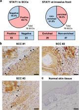
          The process by which cancer cells invade as a cell cluster, known as collective invasion, is associated with metastasis and worse prognosis of cancer patients; therefore, inhibition of collective invasion is considered to improve cancer treatment. However, the cellular characteristics responsible for collective invasion remain largely unknown. Here, we successfully established subclones with various invasive potentials derived from human skin squamous carcinoma cells. The cell cluster of the highly invasive subclone had a hermetically sealed and narrow intercellular space. Interferon-β was localized to the sealed intercellular spaces, leading to collective invasion via the activation of signal transducer and activator of transcription 1 (STAT1). On the other hand, interferon-β was not localized to non-sealed and wide intercellular spaces of the cell cluster of low-invasive subclone with deficient STAT1 activity. In the mixed cell cluster of high- and low-invasive subclones, the high-invasive sub-clonal cells were located at the invasive front of the invasive protrusion, leading to collective invasion by the low-invasive sub-clonal cells. Tissue microarray analysis of human skin squamous cell carcinoma (SCC) also showed enrichment of STAT1 in the invasive front of SCCs. These findings indicate that the intercellular structure controls the potential for collective invasion via STAT1 regulation in SCC.

          Related collections

          Most cited references45

          • Record: found
          • Abstract: found
          • Article: not found

          Regulation of type I interferon responses.

          Type I interferons (IFNs) activate intracellular antimicrobial programmes and influence the development of innate and adaptive immune responses. Canonical type I IFN signalling activates the Janus kinase (JAK)-signal transducer and activator of transcription (STAT) pathway, leading to transcription of IFN-stimulated genes (ISGs). Host, pathogen and environmental factors regulate the responses of cells to this signalling pathway and thus calibrate host defences while limiting tissue damage and preventing autoimmunity. Here, we summarize the signalling and epigenetic mechanisms that regulate type I IFN-induced STAT activation and ISG transcription and translation. These regulatory mechanisms determine the biological outcomes of type I IFN responses and whether pathogens are cleared effectively or chronic infection or autoimmune disease ensues.
            Bookmark
            • Record: found
            • Abstract: found
            • Article: not found

            Circulating tumor cell clusters are oligoclonal precursors of breast cancer metastasis.

            Circulating tumor cell clusters (CTC clusters) are present in the blood of patients with cancer but their contribution to metastasis is not well defined. Using mouse models with tagged mammary tumors, we demonstrate that CTC clusters arise from oligoclonal tumor cell groupings and not from intravascular aggregation events. Although rare in the circulation compared with single CTCs, CTC clusters have 23- to 50-fold increased metastatic potential. In patients with breast cancer, single-cell resolution RNA sequencing of CTC clusters and single CTCs, matched within individual blood samples, identifies the cell junction component plakoglobin as highly differentially expressed. In mouse models, knockdown of plakoglobin abrogates CTC cluster formation and suppresses lung metastases. In breast cancer patients, both abundance of CTC clusters and high tumor plakoglobin levels denote adverse outcomes. Thus, CTC clusters are derived from multicellular groupings of primary tumor cells held together through plakoglobin-dependent intercellular adhesion, and though rare, they greatly contribute to the metastatic spread of cancer. Copyright © 2014 Elsevier Inc. All rights reserved.
              Bookmark
              • Record: found
              • Abstract: found
              • Article: not found

              GSEA-P: a desktop application for Gene Set Enrichment Analysis.

              Gene Set Enrichment Analysis (GSEA) is a computational method that assesses whether an a priori defined set of genes shows statistically significant, concordant differences between two biological states. We report the availability of a new version of the Java based software (GSEA-P 2.0) that represents a major improvement on the previous release through the addition of a leading edge analysis component, seamless integration with the Molecular Signature Database (MSigDB) and an embedded browser that allows users to search for gene sets and map them to a variety of microarray platform formats. This functionality makes it possible for users to directly import gene sets from MSigDB for analysis with GSEA. We have also improved the visualizations in GSEA-P 2.0 and added links to a new form of concise gene set annotations called Gene Set Cards. These additions, as well as other improvements suggested by over 3500 users who have downloaded the software over the past year have been incorporated into this new release of the GSEA-P Java desktop program. GSEA-P 2.0 is freely available for academic and commercial users and can be downloaded from http://www.broad.mit.edu/GSEA
                Bookmark

                Author and article information

                Contributors
                haga@sci.hokudai.ac.jp
                Journal
                Oncogenesis
                Oncogenesis
                Oncogenesis
                Nature Publishing Group UK (London )
                2157-9024
                24 May 2022
                24 May 2022
                December 2022
                : 11
                : 1
                : 27
                Affiliations
                [1 ]GRID grid.39158.36, ISNI 0000 0001 2173 7691, Division of Life Science, Graduate School of Life Science, , Hokkaido University, ; N10-W8, Kita-ku, Sapporo, 060-0810 Japan
                [2 ]GRID grid.39158.36, ISNI 0000 0001 2173 7691, Laboratory of Histology and Cytology, Faculty of Medicine and Graduate School of Medicine, , Hokkaido University, ; N15-W7, Kita-ku, Sapporo, 060-8638 Japan
                [3 ]GRID grid.39158.36, ISNI 0000 0001 2173 7691, Department of Advanced Transdisciplinary Sciences, Faculty of Advanced Life Science, , Hokkaido University, ; N10-W8, Kita-ku, Sapporo, 060-0810 Japan
                [4 ]GRID grid.27476.30, ISNI 0000 0001 0943 978X, Department of Pathology, Graduate School of Medicine, , Nagoya University, ; 65 Tsurumai-cho, Showa-ku, Nagoya, 466-8550 Japan
                [5 ]GRID grid.27476.30, ISNI 0000 0001 0943 978X, Department of Dermatology, , Nagoya University Graduate School of Medicine, ; 65 Tsurumai-cho, Showa-ku, Nagoya, 466-8550 Japan
                Author information
                http://orcid.org/0000-0003-0514-1710
                http://orcid.org/0000-0002-9206-6116
                http://orcid.org/0000-0001-9464-6723
                Article
                403
                10.1038/s41389-022-00403-9
                9126940
                35606369
                73a607e8-5450-45d8-8521-5002f71a4163
                © The Author(s) 2022

                Open Access This article is licensed under a Creative Commons Attribution 4.0 International License, which permits use, sharing, adaptation, distribution and reproduction in any medium or format, as long as you give appropriate credit to the original author(s) and the source, provide a link to the Creative Commons license, and indicate if changes were made. The images or other third party material in this article are included in the article’s Creative Commons license, unless indicated otherwise in a credit line to the material. If material is not included in the article’s Creative Commons license and your intended use is not permitted by statutory regulation or exceeds the permitted use, you will need to obtain permission directly from the copyright holder. To view a copy of this license, visit http://creativecommons.org/licenses/by/4.0/.

                History
                : 17 October 2021
                : 17 April 2022
                : 5 May 2022
                Funding
                Funded by: FundRef https://doi.org/10.13039/100009619, Japan Agency for Medical Research and Development (AMED);
                Award ID: 20gm0810007h0005
                Award Recipient :
                Funded by: FundRef https://doi.org/10.13039/501100001691, MEXT | Japan Society for the Promotion of Science (JSPS);
                Award ID: 21K07142
                Award ID: 20J13568
                Award ID: 21K07141
                Award Recipient :
                Categories
                Article
                Custom metadata
                © The Author(s) 2022

                Oncology & Radiotherapy
                cancer imaging,squamous cell carcinoma,metastasis,cell migration,cell adhesion

                Comments

                Comment on this article