22
views
0
recommends
+1 Recommend
0 collections
    0
    shares
      • Record: found
      • Abstract: found
      • Article: found
      Is Open Access

      Lateral Femoral Bowing and the Location of Atypical Femoral Fractures

      research-article
      , MD, , MD, , MD , , MD
      Hip & Pelvis
      Korean Hip Society
      Femur, Atypical fracture, Lateral bowing angle

      Read this article at

          There is no author summary for this article yet. Authors can add summaries to their articles on ScienceOpen to make them more accessible to a non-specialist audience.

          Abstract

          Purpose

          Atypical femoral fractures (AFFs) occur in two distinct part, subtrochanter and diaphysis. The aim of this study was to investigate the relationship between the lateral femoral bowing angle and the location of AFF.

          Materials and Methods

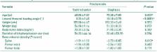
          This study included a total of 56 cases in 45 patients who underwent surgical treatment between January 2010 and December 2015. For the diaphyseal and subtrochanteric AFFs, we evaluated the relationship between the anatomic location and lateral femoral bowing angle. Lateral femoral bowing angle was measured by two orthopaedic surgeons and average value of two calibrators was used in statistic analysis. Other variables like age, height, weight, body mass index and bone mineral density were also evaluated. We also calculated the cutoff value for the location of the fractures from the raw data.

          Results

          The average lateral femoral bowing angle was 10.10°±3.79° (3°-19°) in diaphyseal group and 3.33°±2.45° (1.5°-11°) in subtrochanter group. Lateral femoral bowing angle was statistically significant in logistic regression analysis. According to the receiver operating characteristic curve, cutoff value for the location of the fracture was 5.25°. In other words, the femoral diaphyseal fractures are more frequent if the lateral femoral bowing angle is greater than 5.25°.

          Conclusion

          The lateral femoral bowing angle is associated with the location of the AFFs and the cutoff value of lateral femoral bowing angle was 5.25°.

          Related collections

          Most cited references20

          • Record: found
          • Abstract: found
          • Article: not found

          Atypical subtrochanteric and diaphyseal femoral fractures: second report of a task force of the American Society for Bone and Mineral Research.

          Bisphosphonates (BPs) and denosumab reduce the risk of spine and nonspine fractures. Atypical femur fractures (AFFs) located in the subtrochanteric region and diaphysis of the femur have been reported in patients taking BPs and in patients on denosumab, but they also occur in patients with no exposure to these drugs. In this report, we review studies on the epidemiology, pathogenesis, and medical management of AFFs, published since 2010. This newer evidence suggests that AFFs are stress or insufficiency fractures. The original case definition was revised to highlight radiographic features that distinguish AFFs from ordinary osteoporotic femoral diaphyseal fractures and to provide guidance on the importance of their transverse orientation. The requirement that fractures be noncomminuted was relaxed to include minimal comminution. The periosteal stress reaction at the fracture site was changed from a minor to a major feature. The association with specific diseases and drug exposures was removed from the minor features, because it was considered that these associations should be sought rather than be included in the case definition. Studies with radiographic review consistently report significant associations between AFFs and BP use, although the strength of associations and magnitude of effect vary. Although the relative risk of patients with AFFs taking BPs is high, the absolute risk of AFFs in patients on BPs is low, ranging from 3.2 to 50 cases per 100,000 person-years. However, long-term use may be associated with higher risk (∼100 per 100,000 person-years). BPs localize in areas that are developing stress fractures; suppression of targeted intracortical remodeling at the site of an AFF could impair the processes by which stress fractures normally heal. When BPs are stopped, risk of an AFF may decline. Lower limb geometry and Asian ethnicity may contribute to the risk of AFFs. There is inconsistent evidence that teriparatide may advance healing of AFFs.
            Bookmark
            • Record: found
            • Abstract: found
            • Article: not found

            Severely suppressed bone turnover: a potential complication of alendronate therapy.

            Alendronate, an inhibitor of bone resorption, is widely used in osteoporosis treatment. However, concerns have been raised about potential oversuppression of bone turnover during long-term use. We report on nine patients who sustained spontaneous nonspinal fractures while on alendronate therapy, six of whom displayed either delayed or absent fracture healing for 3 months to 2 yr during therapy. Histomorphometric analysis of the cancellous bone showed markedly suppressed bone formation, with reduced or absent osteoblastic surface in most patients. Osteoclastic surface was low or low-normal in eight patients, and eroded surface was decreased in four. Matrix synthesis was markedly diminished, with absence of double-tetracycline label and absent or reduced single-tetracycline label in all patients. The same trend was seen in the intracortical and endocortical surfaces. Our findings raise the possibility that severe suppression of bone turnover may develop during long-term alendronate therapy, resulting in increased susceptibility to, and delayed healing of, nonspinal fractures. Although coadministration of estrogen or glucocorticoids appears to be a predisposing factor, this apparent complication can also occur with monotherapy. Our observations emphasize the need for increased awareness and monitoring for the potential development of excessive suppression of bone turnover during long-term alendronate therapy.
              Bookmark
              • Record: found
              • Abstract: found
              • Article: not found

              Effects of risedronate treatment on vertebral and nonvertebral fractures in women with postmenopausal osteoporosis: a randomized controlled trial. Vertebral Efficacy With Risedronate Therapy (VERT) Study Group.

              Risedronate, a potent bisphosphonate, has been shown to be effective in the treatment of Paget disease of bone and other metabolic bone diseases but, to our knowledge, it has not been evaluated in the treatment of established postmenopausal osteoporosis. To test the efficacy and safety of daily treatment with risedronate to reduce the risk of vertebral and other fractures in postmenopausal women with established osteoporosis. Randomized, double-blind, placebo-controlled trial of 2458 ambulatory postmenopausal women younger than 85 years with at least 1 vertebral fracture at baseline who were enrolled at 1 of 110 centers in North America conducted between December 1993 and January 1998. Subjects were randomly assigned to receive oral treatment for 3 years with risedronate (2.5 or 5 mg/d) or placebo. All subjects received calcium, 1000 mg/d. Vitamin D (cholecalciferol, up to 500 IU/d) was provided if baseline levels of 25-hydroxyvitamin D were low. Incidence of new vertebral fractures as detected by quantitative and semiquantitative assessments of radiographs; incidence of radiographically confirmed nonvertebral fractures and change from baseline in bone mineral density as determined by dual x-ray absorptiometry. The 2.5 mg/d of risedronate arm was discontinued after 1 year; in the placebo and 5 mg/d of risedronate arms, 450 and 489 subjects, respectively, completed all 3 years of the trial. Treatment with 5 mg/d of risedronate, compared with placebo, decreased the cumulative incidence of new vertebral fractures by 41 % (95% confidence interval [CI], 18%-58%) over 3 years (11.3 % vs 16.3%; P= .003). A fracture reduction of 65% (95% CI, 38%-81 %) was observed after the first year (2.4% vs 6.4%; P<.001). The cumulative incidence of nonvertebral fractures over 3 years was reduced by 39% (95% CI, 6%-61 %) (5.2 % vs 8.4%; P = .02). Bone mineral density increased significantly compared with placebo at the lumbar spine (5.4% vs 1.1 %), femoral neck (1.6% vs -1.2%), femoral trochanter (3.3% vs -0.7%), and midshaft of the radius (0.2% vs -1.4%). Bone formed during risedronate treatment was histologically normal. The overall safety profile of risedronate, including gastrointestinal safety, was similar to that of placebo. These data suggest that risedronate therapy is effective and well tolerated in the treatment of women with established postmenopausal osteoporosis.
                Bookmark

                Author and article information

                Journal
                Hip Pelvis
                Hip Pelvis
                HP
                Hip & Pelvis
                Korean Hip Society
                2287-3260
                2287-3279
                June 2017
                02 June 2017
                : 29
                : 2
                : 127-132
                Affiliations
                Department of Orthopaedic Surgery, Daegu Fatima Hospital, Daegu, Korea.
                Author notes
                Address reprint request to Youngho Cho, MD. Department of Orthopaedic Surgery, Daegu Fatima Hospital, 99 Ayang-ro, Dong-gu, Daegu 41199, Korea. TEL: +82-53-940-7324, FAX: +82-53-954-7417, femur1973@ 123456gmail.com
                Article
                10.5371/hp.2017.29.2.127
                5465394
                599ce4e1-89bb-4b8c-b7ad-d3790e1fb814
                Copyright © 2017 by Korean Hip Society

                This is an Open Access article distributed under the terms of the Creative Commons Attribution Non-Commercial License ( http://creativecommons.org/licenses/by-nc/4.0) which permits unrestricted non-commercial use, distribution, and reproduction in any medium, provided the original work is properly cited.

                History
                : 12 January 2017
                : 11 April 2017
                : 24 April 2017
                Categories
                Original Article

                femur,atypical fracture,lateral bowing angle
                femur, atypical fracture, lateral bowing angle

                Comments

                Comment on this article