0
views
0
recommends
+1 Recommend
0 collections
    0
    shares
      • Record: found
      • Abstract: found
      • Article: found
      Is Open Access

      Intradural view of the spinal cord and dura after three-column osteotomy: illustrative case

      case-report

      Read this article at

          There is no author summary for this article yet. Authors can add summaries to their articles on ScienceOpen to make them more accessible to a non-specialist audience.

          Abstract

          BACKGROUND

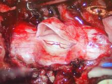
          A three-column osteotomy results in dural buckling, which may appear concerning upon intraoperative visualization because it may appear that the neural elements may also be buckled. The authors presented an intraoperative view after intentional durotomy of the neural elements and the relaxed state of the dura after three-column osteotomy.

          OBSERVATIONS

          A 52-year-old woman with adult tethered cord syndrome and previous untethering presented with worsening leg pain and stiffness, urinary incontinence, and unbalanced gait. Magnetic resonance imaging demonstrated an arachnoid web at T6 and spinal cord tethering. Spinal column shortening via three-column osteotomy was performed with concomitant intradural excision of the arachnoid web. Dural buckling was observed intraoperatively after spinal column shortening. After the durotomy, the spinal cord was visualized without kinking or buckling.

          LESSONS

          Dural buckling after spinal column shortening of 15 mm via three-column osteotomy at T6 did not result in concomitant buckling of the underlying neural elements.

          Related collections

          Most cited references23

          • Record: found
          • Abstract: found
          • Article: not found

          Tethered cord syndrome: an updated review.

          Tethered cord syndrome (TCS) is a diverse clinical entity characterized by symptoms and signs which are caused by excessive tension on the spinal cord. The majority of cases are related to spinal dysraphism. TCS can present in any age group, and presentations differ according to the underlying pathologic condition and age, with pain, cutaneous signs, orthopedic deformities and neurological deficits being the most common. Surgical untethering is indicated in patients with progressive or new onset symptomatology attributable to TCS. The surgical strategy aims to release the tethering structure and thus the chronic tension on the cord. Early operative intervention is associated with improved outcomes. Pain relief is accomplished in almost all cases. Realistic surgical goals include relief of pain and stabilization of neurological function, although improvement in function is often seen. Cord untethering can also halt the progression of scoliosis. The benefits of surgery are debated in asymptomatic patients and patients with normal imaging.
            Bookmark
            • Record: found
            • Abstract: found
            • Article: not found

            Tethered cord syndrome in adults.

            Patients with tethered cord syndrome (TCS) rarely have symptomatic onset in adulthood. Twenty-three adult patients with TCS were studied with respect to the clinical, radiological, and pathological features of this syndrome. Specific circumstances involving either additional tugging of the already tight conus, narrowing of the spinal canal, or direct trauma to the back or buttocks precipitated symptomatic onset in 60% of patients. Diffuse and non-dermatomal leg pain, often referred to the anorectal region, was the most common presenting symptom. Progressive sensorimotor deficits in the lower extremities as well as bladder and bowel dysfunction were also common findings; but, unlike TCS in children, progressive foot and spinal deformities were not seen. As in TCS with onset in childhood, the most common tethering lesions were thickened filum, intradural lipoma, and fibrous adhesions. The degree of cord traction, rather than the type or distribution of the tethering lesions, probably determines the age of symptom onset: less severe traction remains asymptomatic in childhood but results in neurological dysfunction in later life due to repeated tugging of the conus during natural head and neck flexion, or when abnormal tension is aggravated by trauma or spondylotic canal stenosis. Metrizamide myelography revealed the diagnosis of tethered conus in most cases, but the addition of computerized tomographic imaging provided valuable structural details concerning the tethering lesion. The surgical outcome was gratifying in relation to pain and motor weakness but disappointing in the resolution of bowel and bladder dysfunction. Early diagnosis and adequate release of the tethered conus are the keys to successful management.
              Bookmark
              • Record: found
              • Abstract: found
              • Article: not found

              Surgical management of tethered cord syndrome in adults: indications, techniques, and long-term outcomes in 60 patients.

              The adult presentation of tethered cord syndrome (TCS) is well recognized but continues to pose significant diagnostic and management challenges. The authors performed a retrospective study of clinical outcomes after neurosurgical intervention in 60 adults with TCS. All patients who underwent detethering surgery for caudal cord tethering at Toronto Western Hospital between August 1993 and 2004 were identified. Their clinical charts, operative records, and follow-up data were reviewed. Detethering procedures were performed in 62 patients (age range 17-72 years) for TCS of various origins. Long-term (mean 41.5 months) follow-up data were obtained in 60 patients. The tethering lesions were tight terminal filum in 29 patients, postrepair myelomeningocele in 15, lipomyelomeningocele/lipoma in nine, split cord malformation in four, and arachnoidal adhesions in three. Fifty-nine patients presented with progressive pain and/or neurological dysfunction. One patient underwent prophylactic sectioning of the terminal filum. Most patients (71%) had bladder dysfunction at presentation. Microsurgical release of the tethered cord was performed in each case while using multimodality intraoperative neurophysiological monitoring. The most common complication was cerebrospinal fluid leakage, which occurred in nine patients and was managed by reinforcement sutures in four patients, temporary external drainage in three, and the placement of a lumboperitoneal shunt in two. Infective complications included superficial wound infection in three patients, meningitis in one, and urinary tract infection in one. One patient who had undergone multiple previous intradural procedures experienced worsened foot weakness postoperatively. Another patient experienced temporary unilateral lower-limb numbness. At follow up, improvement was noted in the majority of patients presenting with back (78%) and leg (83%) pain. Improvement was more likely in patients with preoperative motor weakness than in those with sensory deficits. Overall, neurological status was improved or stabilized in 90% of patients. Subjective improvement in bladder function was noted in 50% of patients with bladder dysfunction at presentation. Surgery in adult patients with TCS is safe and effective for improving pain and neurological status in the majority of patients; however, patients who have undergone previous intradural detethering procedures in general fare less well, and considerable judgment is required in their management.
                Bookmark

                Author and article information

                Journal
                J Neurosurg Case Lessons
                J Neurosurg Case Lessons
                J Neurosurg Case Lessons
                Journal of Neurosurgery: Case Lessons
                American Association of Neurological Surgeons
                2694-1902
                8 November 2021
                8 November 2021
                : 2
                : 19
                : CASE21497
                Affiliations
                [1 ]Renaissance School of Medicine, Stony Brook University, Stony Brook, New York; and
                [2 ]Department of Neurological Surgery, University of California, San Francisco, San Francisco, California
                Author notes
                Correspondence Zirun Zhao: Renaissance School of Medicine, Stony Brook University, Stony Brook, NY. zirun.zhao@ 123456stonybrookmedicine.edu .

                INCLUDE WHEN CITING Published November 8, 2021; DOI: 10.3171/CASE21497.

                Disclosures Dr. Agarwal reported royalties from Thieme Medical Publishers and Springer International Publishing outside the submitted work. Dr. Mummaneni reported personal fees from Depuy Synthes, Stryker, Globus, Springer International Publishing, and Thieme Publishers outside the submitted work and grants from Neurosurgery Research & Education Foundation (NREF), AO Spine, International Spine Study Group (ISSG), and the National Institutes of Health. Dr. Chou reported personal fees from Globus and Orthofix outside the submitted work.

                Article
                CASE21497
                10.3171/CASE21497
                9435575
                36061976
                fa045da4-66bb-4dd6-862d-b8c2863bc870
                © 2021 The authors

                CC BY-NC-ND 4.0 ( http://creativecommons.org/licenses/by-nc-nd/4.0/).

                History
                : 2 September 2021
                : 1 October 2021
                Page count
                Figures: 3, Tables: 0, References: 23, Pages: 4
                Categories
                Spine, Spine
                Case Lesson

                tethered cord syndrome,spinal column shortening,dural buckling,csf = cerebrospinal fluid,ct = computed tomography,mri = magnetic resonance imaging,tcs = tethered cord syndrome

                Comments

                Comment on this article