17
views
0
recommends
+1 Recommend
0 collections
    0
    shares
      • Record: found
      • Abstract: found
      • Article: found
      Is Open Access

      Glucocorticoids induce senescence in primary human tenocytes by inhibition of sirtuin 1 and activation of the p53/p21 pathway: in vivo and in vitro evidence

      research-article

      Read this article at

          There is no author summary for this article yet. Authors can add summaries to their articles on ScienceOpen to make them more accessible to a non-specialist audience.

          Abstract

          Cellular senescence is an irreversible side effect of some pharmaceuticals which can contribute to tissue degeneration.

          Objective

          To determine whether pharmaceutical glucocorticoids induce senescence in tenocytes.

          Methods

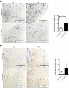
          Features of senescence (β-galactosidase activity at pH 6 (SA-β-gal) and active mammalian/mechanistic target of rapamycin (mTOR) in cell cycle arrest) as well as the activity of the two main pathways leading to cell senescence were examined in glucocorticoid-treated primary human tenocytes. Evidence of senescence-inducing pathway induction in vivo was obtained using immunohistochemistry on tendon biopsy specimens taken before and 7 weeks after subacromial Depo-Medrone injection.

          Results

          Dexamethasone treatment of tenocytes resulted in an increased percentage of SA-βgal-positive cells. Levels of phosphorylated p70S6K did not decrease with glucocorticoid treatment indicating mTOR remained active. Increased levels of acetylated p53 as well as increased RNA levels of its pro-senescence effector p21 were evident in dexamethasone-treated tenocytes. Levels of the p53 deacetylase sirtuin 1 were lower in dexamethasone-treated cells compared with controls. Knockdown of p53 or inhibition of p53 activity prevented dexamethasone-induced senescence. Activation of sirtuin 1 either by exogenous overexpression or by treatment with resveratrol or low glucose prevented dexamethasone-induced senescence. Immunohistochemical analysis of tendon biopsies taken before and after glucocorticoid injection revealed a significant increase in the percentage of p53-positive cells (p=0.03). The percentage of p21-positive cells also tended to be higher post-injection (p=0.06) suggesting glucocorticoids activate the p53/p21 senescence-inducing pathway in vivo as well as in vitro.

          Conclusion

          As cell senescence is irreversible in vivo, glucocorticoid-induced senescence may result in long-term degenerative changes in tendon tissue.

          Related collections

          Most cited references38

          • Record: found
          • Abstract: found
          • Article: not found

          Negative control of p53 by Sir2alpha promotes cell survival under stress.

          The NAD-dependent histone deacetylation of Sir2 connects cellular metabolism with gene silencing as well as aging in yeast. Here, we show that mammalian Sir2alpha physically interacts with p53 and attenuates p53-mediated functions. Nicotinamide (Vitamin B3) inhibits an NAD-dependent p53 deacetylation induced by Sir2alpha, and also enhances the p53 acetylation levels in vivo. Furthermore, Sir2alpha represses p53-dependent apoptosis in response to DNA damage and oxidative stress, whereas expression of a Sir2alpha point mutant increases the sensitivity of cells in the stress response. Thus, our findings implicate a p53 regulatory pathway mediated by mammalian Sir2alpha. These results have significant implications regarding an important role for Sir2alpha in modulating the sensitivity of cells in p53-dependent apoptotic response and the possible effect in cancer therapy.
            Bookmark
            • Record: found
            • Abstract: found
            • Article: not found

            Human SIR2 deacetylates p53 and antagonizes PML/p53-induced cellular senescence.

            The yeast Sir2 protein mediates chromatin silencing through an intrinsic NAD-dependent histone deacetylase activity. Sir2 is a conserved protein and was recently shown to regulate lifespan extension both in budding yeast and worms. Here, we show that SIRT1, the human Sir2 homolog, is recruited to the promyelocytic leukemia protein (PML) nuclear bodies of mammalian cells upon overexpression of either PML or oncogenic Ras (Ha-rasV12). SIRT1 binds and deacetylates p53, a component of PML nuclear bodies, and it can repress p53-mediated transactivation. Moreover, we show that SIRT1 and p53 co-localize in nuclear bodies upon PML upregulation. When overexpressed in primary mouse embryo fibroblasts (MEFs), SIRT1 antagonizes PML-induced acetylation of p53 and rescues PML-mediated premature cellular senescence. Taken together, our data establish the SIRT1 deacetylase as a novel negative regulator of p53 function capable of modulating cellular senescence.
              Bookmark
              • Record: found
              • Abstract: found
              • Article: not found

              Nutrient availability regulates SIRT1 through a forkhead-dependent pathway.

              Nutrient availability regulates life-span in a wide range of organisms. We demonstrate that in mammalian cells, acute nutrient withdrawal simultaneously augments expression of the SIRT1 deacetylase and activates the Forkhead transcription factor Foxo3a. Knockdown of Foxo3a expression inhibited the starvation-induced increase in SIRT1 expression. Stimulation of SIRT1 transcription by Foxo3a was mediated through two p53 binding sites present in the SIRT1 promoter, and a nutrient-sensitive physical interaction was observed between Foxo3a and p53. SIRT1 expression was not induced in starved p53-deficient mice. Thus, in mammalian cells, p53, Foxo3a, and SIRT1, three proteins separately implicated in aging, constitute a nutrient-sensing pathway.
                Bookmark

                Author and article information

                Journal
                Ann Rheum Dis
                Ann. Rheum. Dis
                annrheumdis
                ard
                Annals of the Rheumatic Diseases
                BMJ Publishing Group (BMA House, Tavistock Square, London, WC1H 9JR )
                0003-4967
                1468-2060
                July 2014
                1 June 2013
                : 73
                : 7
                : 1405-1413
                Affiliations
                Nuffield Department of Orthopaedics, Rheumatology and Musculoskeletal Sciences, Botnar Research Centre, University of Oxford , Oxford, UK
                Author notes

                Handling editor Tore K Kvien

                [Correspondence to ] Dr Raewyn C Poulsen, Nuffield Department of Orthopaedics, Rheumatology and Musculoskeletal Sciences, Botnar Research Centre, University of Oxford, Oxford OX3 7D, UK; raewyn.poulsen@ 123456ndorms.ox.ac.uk
                Article
                annrheumdis-2012-203146
                10.1136/annrheumdis-2012-203146
                4078757
                23727633
                6eb69f65-230c-47dd-82e8-97ff12cb5c48
                Published by the BMJ Publishing Group Limited. For permission to use (where not already granted under a licence) please go to http://group.bmj.com/group/rights-licensing/permissions

                This is an Open Access article distributed in accordance with the Creative Commons Attribution Non Commercial (CC BY-NC 3.0) license, which permits others to distribute, remix, adapt, build upon this work non-commercially, and license their derivative works on different terms, provided the original work is properly cited and the use is non-commercial. See: http://creativecommons.org/licenses/by-nc/3.0/

                History
                : 7 May 2013
                Categories
                1506
                Basic and Translational Research
                Extended Report
                Custom metadata
                unlocked

                Immunology
                corticosteroids,tendinitis,chondrocytes,fibroblasts,inflammation
                Immunology
                corticosteroids, tendinitis, chondrocytes, fibroblasts, inflammation

                Comments

                Comment on this article