1
views
0
recommends
+1 Recommend
0 collections
    0
    shares
      • Record: found
      • Abstract: found
      • Article: found
      Is Open Access

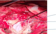
      Contiguous diastematomyelia with tethered cord, intradural extramedullary dermoid tumor, and lipomyelomeningocele: A unique case of spinal dysraphism

      case-report

      Read this article at

          There is no author summary for this article yet. Authors can add summaries to their articles on ScienceOpen to make them more accessible to a non-specialist audience.

          Key Clinical Message

          Diastematomyelia, tethered cord, intradural extramedullary dermoid tumor and lipomyelomeningocele such disease entities themselves are rare in their own form and concurrent presentation of all those pathological states in a single individual can be considered one of the rarest forms of spinal dysraphism globally. Moreover for prompt management with optimal prognosis needs refined neurosurgical intervention guided by intraoperative neuromonitoring so as to bring about the best quality of life in the patient.

          Related collections

          Most cited references10

          • Record: found
          • Abstract: found
          • Article: not found

          Management of lipomyelomeningoceles. Experience at the Hospital for Sick Children, Toronto.

          Ninety-seven children with lipomyelomeningoceles were operated on at the Hospital for Sick Children between January, 1960, and December, 1982. The most common factor that caused these patients to seek help was the cosmetic effect of the mass on their back. However, 22 patients had urinary incontinence and 15 patients had a deformed or weak leg. Sixty of the 97 patients were female and 37 were male. The patients presented for treatment between 6 days and 18 1/2 years of age with a median age of 7 1/2 months. Fifty-six patients presented before the age of 6 months and 35 of these were perfectly normal at the time of presentation. On the other hand, of the 41 patients who were brought for treatment after the age of 6 months, only 12 were normal prior to surgery. When patients were appropriately treated at an early age, with their spinal cords untethered and their dura securely closed with a dural graft, then they remained unchanged neurologically or even improved. However, when treatment was delayed or not done appropriately then they were left with significant neurological sequelae. Lipomyelomeningoceles are serious lesions which without appropriate therapy can result in gross impairment of neurological function.
            Bookmark
            • Record: found
            • Abstract: found
            • Article: not found

            Intramedullary Masses of the Spinal Cord: Radiologic-Pathologic Correlation.

            Spinal cord tumors are a challenge for patients and neurosurgeons because of the high risk of neurologic deficits from the disease process and surgical interventions. Spinal cord tumors are uncommon, and approximately 2%-3% of primary intra-axial tumors of the central nervous system occur in the spinal cord. Primary intra-axial tumors are usually derived from neuroepithelial tissue, especially glial cells. This often leads to a classic intramedullary mass differential diagnosis of ependymoma or astrocytoma, which together constitute up to 70% of spinal cord tumors. For example, ependymomas occur predominantly in adults, and astrocytomas (specifically pilocytic astrocytomas) occur predominantly in children. While that is an excellent starting point, in order to refine the differential diagnosis, the authors review the radiologic-pathologic features of specific neoplastic categories and entities recognized by the World Health Organization (WHO) in the 2016 WHO Classification of Tumours of the Central Nervous System and a few additional congenital-developmental entities. Radiologists can add value by providing a reasonable preoperative differential diagnosis for the patient and neurosurgeon, in many cases by favoring the most common conditions, and in other cases by identifying radiologic features that may point toward a less common entity. Some of the less common entities include intramedullary myxopapillary ependymoma, spinal subependymoma, and spinal hemangioblastoma. Whenever possible, the characteristic imaging features and locations of these tumors are explained or traced back to the underlying cell of origin and findings seen at histopathologic examination.See discussion on this article by Buch.
              Bookmark
              • Record: found
              • Abstract: found
              • Article: not found

              Lipomyelomeningocele: pathology, treatment, and outcomes.

              Lipomyelomeningocele represents a rare but complex neurological disorder that may present with neurological deterioration secondary to an inherent tethered spinal cord. Radiological testing is beneficial in determining the morphology of the malformation. Specialized testing such as urodynamic studies and neurophysiological testing may be beneficial in assessing for neurological dysfunction secondary to the lipomyelomeningocele. Early surgical intervention may be beneficial in preventing further neurological decline.
                Bookmark

                Author and article information

                Contributors
                sagunghimire01@gmail.com
                Journal
                Clin Case Rep
                Clin Case Rep
                10.1002/(ISSN)2050-0904
                CCR3
                Clinical Case Reports
                John Wiley and Sons Inc. (Hoboken )
                2050-0904
                20 April 2024
                April 2024
                : 12
                : 4 ( doiID: 10.1002/ccr3.v12.4 )
                : e8783
                Affiliations
                [ 1 ] Department of Neurosurgery B and B Hospital Lalitpur Nepal
                [ 2 ] Department of Radiology B and B Hospital Lalitpur Nepal
                [ 3 ] Department of Neuroscience B and B Hospital Lalitpur Nepal
                [ 4 ] KIST Medical College and Teaching Hospital Lalitpur Nepal
                Author notes
                [*] [* ] Correspondence

                Sagun Ghimire, Department of Neurosurgery, B and B Hospital, Gwarko, Lalitpur, Nepal.

                Email: sagunghimire01@ 123456gmail.com

                Author information
                https://orcid.org/0000-0001-5195-1998
                https://orcid.org/0009-0006-7156-7604
                Article
                CCR38783 CCR3-2024-03-0749.R1
                10.1002/ccr3.8783
                11031741
                38645607
                ab4a1ca4-cefd-40aa-89e1-5e46d1e86a4f
                © 2024 The Authors. Clinical Case Reports published by John Wiley & Sons Ltd.

                This is an open access article under the terms of the http://creativecommons.org/licenses/by-nc-nd/4.0/ License, which permits use and distribution in any medium, provided the original work is properly cited, the use is non‐commercial and no modifications or adaptations are made.

                History
                : 26 March 2024
                : 16 March 2024
                : 29 March 2024
                Page count
                Figures: 6, Tables: 0, Pages: 5, Words: 2400
                Categories
                Neurology
                Neurosurgery
                Radiology & Imaging
                Surgery
                Case Report
                Case Report
                Custom metadata
                2.0
                April 2024
                Converter:WILEY_ML3GV2_TO_JATSPMC version:6.4.0 mode:remove_FC converted:20.04.2024

                diastematomyelia,lipomyelomeningocele,spinal dysraphism,tethered cord

                Comments

                Comment on this article