39
views
0
recommends
+1 Recommend
0 collections
    0
    shares
      • Record: found
      • Abstract: found
      • Article: found
      Is Open Access

      Transcranial doppler sonography is not a valid diagnostic tool for detection of basilar artery stenosis or in-stent restenosis: a retrospective diagnostic study

      research-article

      Read this article at

          There is no author summary for this article yet. Authors can add summaries to their articles on ScienceOpen to make them more accessible to a non-specialist audience.

          Abstract

          Background

          There are contradictory reports concerning the validity of transcranial sonography (TCD and TCCS) for examinations of the basilar artery. Here we investigated sensitivity and specificity of transcranial sonography for the detection of basilar artery stenosis and in-stent-restenosis compared to cerebral angiography.

          Methods

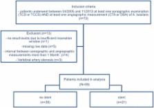
          We analyzed data of 104 examinations of the basilar artery. The association between sonographic peak systolic velocity (PSV) and degree of stenosis obtained by cerebral angiography was evaluated applying Spearman’s correlation coefficient. Receiver Operating Characteristics (ROC) curves and areas under the curve (AUC) were calculated for the detection of a ≥50% stenosis defined by angiography. Optimal cut-off was derived using the Youden-index.

          Results

          A weak but statistically significant correlation between PSV and the degree of stenosis was found ( n=104, rho=0.35, p<0.001). ROC analysis for a detection of ≥50% stenosis showed an AUC of 0.70, a sensitivity of 74.0% and a specificity of 65.0% at the optimal cut off of 124 cm/s. Results were consistent when analyzing examinations done in stented and unstented arteries separately (TCD VS DSA/CTA in unstented artery: AUC=0.66, sensitivity 61.0%, specificity 65.0%, TCD/TCCS VS DSA in stented artery: AUC=0.63, sensitivity 71.0%, specificity 82.0%). Comparing TCCS measurements exclusively to angiography, ROC analysis showed an AUC of 1.00 for the detection of an in-stent-restenosis ≥50% with a sensitivity and specificity of 100% when a PSV of 132 cm/s was used as a cut off value.

          Conclusion

          Validity of TCD in the assessment of basilar artery stenosis or in-stent restenosis is poor. First results for TCCS are promising, but due to the small samplesize further studies with larger samples sizes are warranted.

          Related collections

          Most cited references23

          • Record: found
          • Abstract: found
          • Article: found

          Twisted Blood Vessels: Symptoms, Etiology and Biomechanical Mechanisms

          Tortuous arteries and veins are commonly observed in humans and animals. While mild tortuosity is asymptomatic, severe tortuosity can lead to ischemic attack in distal organs. Clinical observations have linked tortuous arteries and veins with aging, atherosclerosis, hypertension, genetic defects and diabetes mellitus. However, the mechanisms of their formation and development are poorly understood. This review summarizes the current clinical and biomechanical studies on the initiation, development and treatment of tortuous blood vessels. We submit a new hypothesis that mechanical instability and remodeling could be mechanisms for the initiation and development of these tortuous vessels.
            Bookmark
            • Record: found
            • Abstract: found
            • Article: not found

            Intracranial vascular stenosis and occlusive disease: evaluation with CT angiography, MR angiography, and digital subtraction angiography.

            Although digital subtraction angiography (DSA) provides excellent visualization of the intracranial vasculature, it has several limitations. Our purpose was to evaluate the ability of helical CT angiography (CTA) to help detect and quantify intracranial stenosis and occlusion compared with DSA and MR angiography (MRA). Twenty-eight patients underwent CTA, DSA, and 3D time-of-flight (TOF) MRA for suspected cerebrovascular lesions. All three studies were performed within a 30-day period. Two readers blinded to prior estimated or calculated stenoses, patient history and clinical information examined 672 vessel segments. Lesions were categorized as normal (0-9%), mild (10-29%), moderate (30-69%), severe (70-99%), or occluded (no flow detected). DSA was the reference standard. Unblinded consensus readings were obtained for all discrepancies. A total of 115 diseased vessel segments were identified. After consensus interpretation, CTA revealed higher sensitivity than that of MRA for intracranial stenosis (98% versus 70%, P < .001) and occlusion (100% versus 87%, P = .02). CTA had a higher positive predictive value than that of MRA for both stenosis (93% versus 65%, P < .001) and occlusion (100% versus 59%, P < .001). CTA had a high interoperator reliability. In 6 of 28 patients (21%), all 6 with low-flow states in the posterior circulation, CTA was superior to DSA in detection of vessel patency. CTA has a higher sensitivity and positive predictive value than MRA and is recommended over TOF MRA for detection of intracranial stenosis and occlusion. CTA has a high interoperator reliability. CTA is superior to DSA in the evaluation of posterior circulation steno-occlusive disease when slow flow is present. CTA results had a significant effect on patient clinical management.
              Bookmark
              • Record: found
              • Abstract: found
              • Article: not found

              How accurate is CT angiography in evaluating intracranial atherosclerotic disease?

              Digital subtraction angiography (DSA) is regarded as the gold standard in assessing degree of stenosis in intracranial vessels. However, it is invasive and can only be carried out at specialized centers. We sought to compare CT angiography (CTA) to DSA for detection and measurement of stenosis in large intracranial arteries. We identified all subjects admitted with ischemic stroke or transient ischemic attack and with CTA and DSA studies of good quality completed within 30 days of each other between April 2000 and May 2006 at a single medical center. Two readers blinded to clinical information reviewed each CTA and DSA independently. Each reader located and measured stenosis of 15 prespecified large intracranial arterial segments per study at the same level of magnification. These stenotic lesions were most likely atherosclerotic in etiology. All measurements were made with Wiha digiMax 6" digital calipers. The degree of stenosis was calculated using the published method for the Warfarin-Aspirin Symptomatic Intracranial Disease study. All disagreements of greater than 10% were reviewed by a third reader who decided between the 2 prior measurements. Segments were excluded from analyses if they were judged to be congenitally hypoplastic or seen only through collaterals or cross-filling. Intraclass correlation, sensitivity, and specificity were calculated using DSA as the reference standard. Forty-one pairs of CTA and DSAs from 41 patients were reviewed. CTAs were completed within 28 days before 13 days after DSA, with a median of 1 day. A total of 475 pairs of major intracranial arterial segment were analyzed. Intraclass correlation between degree of stenosis based on CTA and DSA for all segments was 0.98 (P=0.001). CTA detected large arterial occlusion with 100% sensitivity and specificity. For detection of >or=50% stenosis, CTA had 97.1% sensitivity and 99.5% specificity. To detect all lesions >or=50% as determined by DSA, the cut off point on CTA appeared to be at >or=30%, with a false-positive rate of 2.4%. Compared to DSA, CTA has high sensitivity and specificity for detecting >or=50% stenosis of large intracranial arterial segments. CTA is minimally invasive and may be a useful screening tool for intracranial arterial disease and occlusion.
                Bookmark

                Author and article information

                Contributors
                +49-551-3966951 , jliman@gwdg.de
                Journal
                BMC Neurol
                BMC Neurol
                BMC Neurology
                BioMed Central (London )
                1471-2377
                11 May 2017
                11 May 2017
                2017
                : 17
                : 89
                Affiliations
                [1 ]ISNI 0000 0001 2364 4210, GRID grid.7450.6, Department of Neurology, , University of Goettingen, ; Robert-Koch-Str. 40, 37075 Goettingen, Germany
                [2 ]ISNI 0000 0001 2364 4210, GRID grid.7450.6, Department of Neuroradiology, , University of Goettingen, ; Robert-Koch-Str. 40, 37075 Goettingen, Germany
                [3 ]GRID grid.7490.a, Epidemiological and Statistical Methods, Department of Epidemiology, , Helmholtz Centre for Infection Research, ; Inhoffenstr. 7, 38124 Braunschweig, Germany
                Author information
                http://orcid.org/0000-0002-7465-9655
                Article
                872
                10.1186/s12883-017-0872-8
                5426050
                2369e2fc-67ec-4131-be88-c218abb61d65
                © The Author(s). 2017

                Open AccessThis article is distributed under the terms of the Creative Commons Attribution 4.0 International License ( http://creativecommons.org/licenses/by/4.0/), which permits unrestricted use, distribution, and reproduction in any medium, provided you give appropriate credit to the original author(s) and the source, provide a link to the Creative Commons license, and indicate if changes were made. The Creative Commons Public Domain Dedication waiver ( http://creativecommons.org/publicdomain/zero/1.0/) applies to the data made available in this article, unless otherwise stated.

                History
                : 5 October 2016
                : 6 May 2017
                Categories
                Research Article
                Custom metadata
                © The Author(s) 2017

                Neurology
                doppler sonography,duplex sonography,basilar artery stenosis,in stent restenosis
                Neurology
                doppler sonography, duplex sonography, basilar artery stenosis, in stent restenosis

                Comments

                Comment on this article