48
views
0
recommends
+1 Recommend
0 collections
    16
    shares
      • Record: found
      • Abstract: found
      • Article: not found

      Phosphorylation of the Kinase Domain Regulates Autophosphorylation of Myosin IIIA and Its Translocation in Microvilli

      research-article

      Read this article at

      ScienceOpenPublisherPMC
          There is no author summary for this article yet. Authors can add summaries to their articles on ScienceOpen to make them more accessible to a non-specialist audience.

          Abstract

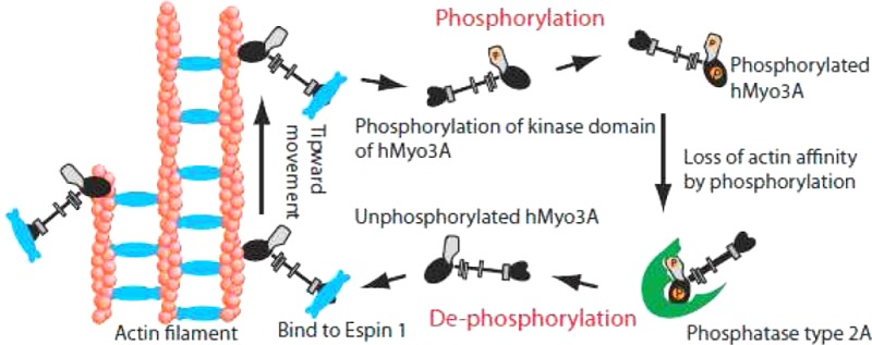
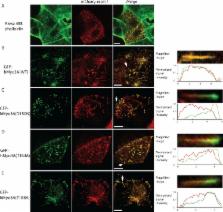
          Motor activity of myosin III is regulated by autophosphorylation. To investigate the role of the kinase activity on the transporter function of myosin IIIA (Myo3A), we identified the phosphorylation sites of kinase domain (KD), which is responsible for the regulation of kinase activity and thus motor function. Using mass spectrometry, we identified six phosphorylation sites in the KD, which are highly conserved among class III myosins and Ste20-related misshapen (Msn) kinases. Two predominant sites, Thr 184 and Thr 188, in KD are important for phosphorylation of the KD as well as the motor domain, which regulates the affinity for actin. In the Caco2 cells, the full-length human Myo3A (hMyo3AFull) markedly enlarged the microvilli, although it did not show discrete localization within the microvilli. On the other hand, hMyo3AFull(T184A) and hMyo3AFull(T188A) both showed clear localization at the microvilli tips. Our results suggest that Myo3A induces large actin bundle formation to form microvilli, and phosphorylation of KD at Thr 184 and Thr 188 is critical for the kinase activity of Myo3A, and regulation of Myo3A translocation to the tip of microvilli. Retinal extracts potently dephosphorylate both KD and motor domain without IQ motifs (MDIQo), which was inhibited by okadaic acid (OA) with nanomolar range and by tautomycetin (TMC) with micromolar range. The results suggest that Myo3A phosphatase is protein phosphatase type 2A (PP2A). Supporting this result, recombinant PP2Ac potently dephosphorylates both KD and MDIQo. We propose that the phosphorylation–dephosphorylation mechanism plays an essential role in mediating the transport and actin bundle formation and stability functions of hMyo3A.

          Related collections

          Most cited references36

          • Record: found
          • Abstract: found
          • Article: not found

          The protein kinase family: conserved features and deduced phylogeny of the catalytic domains.

          In recent years, members of the protein kinase family have been discovered at an accelerated pace. Most were first described, not through the traditional biochemical approach of protein purification and enzyme assay, but as putative protein kinase amino acid sequences deduced from the nucleotide sequences of molecularly cloned genes or complementary DNAs. Phylogenetic mapping of the conserved protein kinase catalytic domains can serve as a useful first step in the functional characterization of these newly identified family members.
            Bookmark
            • Record: found
            • Abstract: found
            • Article: not found

            Site-directed mutagenesis of double-stranded DNA by the polymerase chain reaction.

            We have developed a facile procedure for rapid PCR-based site-directed mutagenesis of double-stranded DNA. Increasing the initial template concentration and decreasing the PCR cycles to 5-10 allows us to reduce the rate of undesired second-site mutations and dramatically increase the time savings. Following PCR, DpnI treatment is used to select against parental DNA molecules. The DpnI (target sequence 5'-Gm6ATC) is specific for methylated and hemimethylated DNA and is used to digest parental DNA and select for mutation-containing amplified DNA. DNA isolated from almost all common Escherichia coli strains is Dam methylated and therefore susceptible to DpnI digestion. Pfu DNA polymerase is used, prior to intramolecular ligation of the linear template, to remove any bases extended onto the 3' ends of the PCR product by Taq DNA polymerase. The recircularized vector DNA incorporating the desired mutations is transformed into E. coli. This method can be used independently of any host strain and vector.
              Bookmark
              • Record: found
              • Abstract: found
              • Article: not found

              From flies' eyes to our ears: mutations in a human class III myosin cause progressive nonsyndromic hearing loss DFNB30.

              Normal vision in Drosophila requires NINAC, a class III myosin. Class III myosins are hybrid motor-signaling molecules, with an N-terminal kinase domain, highly conserved head and neck domains, and a class III-specific tail domain. In Drosophila rhabdomeres, NINAC interacts with actin filaments and with a PDZ scaffolding protein to organize the phototransduction machinery into a signaling complex. Recessive null mutations in Drosophila NINAC delay termination of the photoreceptor response and lead to progressive retinal degeneration. Here, we show that normal hearing in humans requires myosin IIIA, the human homolog of NINAC. In an extended Israeli family, nonsyndromic progressive hearing loss is caused by three different recessive, loss-of-function mutations in myosin IIIA. Of 18 affected relatives in Family N, 7 are homozygous and 11 are compound heterozygous for pairs of mutant alleles. Expression of mammalian myosin IIIA is highly restricted, with the strongest expression in retina and cochlea. The involvement of homologous class III myosins in both Drosophila vision and human hearing is an evolutionary link between these sensory systems.
                Bookmark

                Author and article information

                Journal
                Biochemistry
                Biochemistry
                bi
                bichaw
                Biochemistry
                American Chemical Society
                0006-2960
                1520-4995
                17 November 2015
                17 November 2014
                16 December 2014
                : 53
                : 49
                : 7835-7845
                Affiliations
                []Department of Microbiology and Physiological Systems, University of Massachusetts Medical School , Worcester, Massachusetts 01605, United States
                []Department of Cellular and Molecular Biology, University of Texas Health Science Center at Tyler , Tyler, Texas75708, United States
                [§ ]Department of Molecular Physiology and Medical Bioregulation, Yamaguchi University Graduate School of Medicine , Ube 755-8505, Japan
                []Division of Mass Spectrometry, Korea Basic Science Institute , Ochang-eup, Cheongwon-gun, Chungbuk 363-883, Korea
                Author notes
                [* ]Mitsuo Ikebe. Tel: (903) 877-7785. Fax: (903) 877-5438. E-mail: Mitsuo.Ikebe@ 123456uthct.edu .
                Article
                10.1021/bi501247z
                4270376
                25402663
                8ca0c5ab-5d1f-4b1a-a51a-ad5a81e8c1eb
                Copyright © 2014 American Chemical Society

                This is an open access article published under an ACS AuthorChoice License, which permits copying and redistribution of the article or any adaptations for non-commercial purposes.

                History
                : 02 October 2014
                : 14 November 2014
                Funding
                National Institutes of Health, United States
                Categories
                Article
                Custom metadata
                bi501247z
                bi-2014-01247z

                Biochemistry
                Biochemistry

                Comments

                Comment on this article