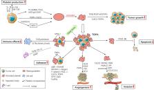
Glioblastoma tumor cells release microvesicles (exosomes) containing mRNA, miRNA and angiogenic proteins. These microvesicles are taken up by normal host cells, such as brain microvascular endothelial cells. By incorporating an mRNA for a reporter protein into these microvesicles we demonstrate that microvesicle-delivered messages are translated by recipient cells. These microvesicles are also enriched in angiogenic proteins and elicit tubule formation by endothelial cells. Tumor-derived microvesicles therefore serve as a novel means of delivery of genetic information as well as proteins to recipient cells in the tumor environment. Glioblastoma microvesicles also stimulated proliferation of a human glioma cell line, indicating a self-promoting aspect. Messenger RNA mutant/variants and microRNAs characteristic of gliomas can be detected in serum microvesicles of glioblastoma patients. The tumor-specific EGFRvIII was detected in serum microvesicles from 7 out of 25 glioblastoma patients. Thus, tumor-derived microvesicles may provide diagnostic information and aid in therapeutic decisions for cancer patients through a blood test. Glioblastomas are highly malignant brain tumors with a poor prognosis despite intensive research and clinical efforts1. These tumors as well as many others have a remarkable ability to mold their stromal environment to their own advantage. Tumor cells alter surrounding normal cells to facilitate tumor cell growth, invasion, chemoresistance, immune evasion and metastasis 2–4. The tumor cells also hijack the normal vasculature and stimulate rapid formation of new blood vessels to supply tumor nutrition 5. Although the immune system can initially suppress tumor growth, it is often progressively blunted by tumor activation of immunosuppressive pathways 6. Recent studies show the importance of communication between tumor cells and their environment through shedding of membrane microvesicles which can fuse to cells in the vicinity 7. Microvesicles are 30–100 nm in diameter and shed from many different cell types under both normal and pathological conditions 8. These exosomes can be formed through inward budding of endosomal membranes giving rise to intracellular multivesicular bodies (MVB) that later fuse with the plasma membrane, releasing the exosomes to the exterior 8,9. They can also be shed directly by outward budding of the plasma membrane, as shown for Jurkat T-cells 10. Microvesicles in Drosophila, termed argosomes, contain morphogens such as Wingless protein and move throughout the imaginal disc epithelium in the developing embryos 11. Microvesicles found in semen, known as prostasomes, can promote sperm motility, stabilize the acrosome reaction, facilitate immunosuppression and inhibit angiogenesis 12. On the other hand, prostasomes released by malignant prostate cells promote angiogenesis. It has been shown that microvesicles can transfer some of their contents to other cell types 13–16. The content of microvesicles and their biological function depends on the cell of origin. Microvesicles derived from B-cells and dendritic cells have potent immuno-stimulatory and antitumor effects in vivo and have been used as antitumor vaccines 17. Dendritic cell-derived microvesicles contain co-stimulatory proteins necessary for T-cell activation, whereas most tumor cell-derived microvesicles do not. Instead they act to suppress the immune response and accelerate tumor growth and invasiveness 18–21. Breast cancer microvesicles stimulate angiogenesis, and platelet-derived microvesicles promote tumor progression and metastasis of lung cancer cells 22,23. Human glioblastoma tissues were obtained from surgical resections and tumor cells were dissociated and cultured as monolayers in medium using fetal bovine serum (FBS) depleted for microvesicles (dFBS). Cultured primary cells obtained from three glioblastoma tumors were found to produce microvesicles at early and later passages (1–15 passages). Tumor cells were covered with microvesicles varying in size from about 50 – 500 nm (Fig. 1a and b). The microvesicles contained RNA and protein in an approximate ratio of 1:80. To evaluate whether the RNA was contained inside the microvesicles, they were either exposed to RNase A or left untreated before RNA extraction (Fig. 1c). There was always less than a 7% decrease in RNA content following RNase treatment. Thus, it appears that almost all of the RNA is contained within the vesicles and is thereby protected from external RNases by the surrounding membrane. Bioanalysis of RNA from microvesicles and their donor cells revealed that the microvesicles contain a broad range of RNA sizes consistent with a variety of mRNAs and miRNAs, but lack the ribosomal RNA peaks characteristic of cellular RNA (Fig. 1d and e). Microarray analysis of mRNA populations in microvesicles and their donor glioblastoma cells was performed using the Agilent 44K whole genome microarray. Approximately 22,000 gene transcripts were found in the cells and 27,000 transcripts in the microvesicles (detected at well above background levels, 99% confidence interval) on both arrays. Approximately 4,700 different mRNAs were detected exclusively in microvesicles on both arrays, indicating a selective enrichment process within the microvesicles (Supplementary Table 1). Consistent with this, there was a poor overall correlation in levels of mRNAs in the cells as compared to microvesicles from two tumor preparations (Fig. 2a and b), supporting selective enrichment of some cellular mRNAs in microvesicles. In contrast, a comparison of levels of specific mRNAs in different preparations of donor cells or of microvesicles showed a strong correlation, indicating a consistent distribution within these distinct cellular compartments (Fig. 2c and d). We found 3426 transcripts differentially distributed more than 5-fold (p-value 95%. The cells were stably transduced and microvesicles generated during the subsequent passages (2–10) were isolated and purified as above. Microvesicles (50 μg) were added to 50,000 HBMVEC and incubated for 24 hrs. The Gluc activity in the supernatant was measured directly after microvesicle addition (0 hrs), and 15 hrs and 24 hrs later and normalised to the Gluc activity in the microvesicles. The results are presented as the mean ± SEM (n = 4). PKH67 labelled microvesicle Purified glioblastoma microvesicles were labelled with PKH67 Green Fluorescent labelling kit (Sigma-Aldrich, St Louis, MO, USA) as described 21. The labelled microvesicles were incubated with HBMVEC in culture (5 μg/50,000 cells). Microvesicles were allowed to bind for 20 min at 4°C and cells were then washed and incubated at 37°C for 1 hr. RT PCR and nested PCR RNA was extracted using the MirVana RNA isolation kit. RNA was converted to cDNA using the Omniscript RT kit (if starting material was >50 ng) or Sensiscript RT kit (if starting material was <50 ng) (Qiagen Inc., Valencia, CA, USA) using a mix of oligo dT and random hexamer primer according to manufacturer’s recommendation. The following PCR primers were used: GAPDH primers; Forw 5′-GAA GGT GAA GGT CGG AGT C-3′, Reverse 5′-GAA GAT GGT GAT GGG ATT TC-3′. EGFR/EGFRvIII PCR1; Forw 5′-CCAGTATTGATCGGGAGAGC-3′, Reverse 5′-TCAGAATATCCAGTTCCTGTGG-3′, EGFR/EGFRvIII PCR2; Forw 5′-ATG CGA CCC TCC GGG ACG-3′, Reverse 5′-GAG TAT GTG TGA AGG AGT-3′. The Gluc primers have been described previously 24. PCR protocol: 94°C 3 min; 94°C 45 s, 60°C 45 s, 72°C 2 min × 35 cycles; 72°C 7 min. Angiogenesis antibody array One mg total protein from either primary glioblastoma cells or purified microvesicles isolated from the same cells were lysed in Promega lysis buffer (Promega, Madison, WI, USA) and then added to the human angiogenesis antibody array (Panomics, Fremont, CA, USA) according to manufacturer’s recommendations. The arrays were scanned and analysed with the ImageJ software (NIH). Statistics The statistical analyses were performed using Students t-test. Supplementary Material 1