11
views
0
recommends
+1 Recommend
0 collections
    0
    shares
      • Record: found
      • Abstract: found
      • Article: found
      Is Open Access

      Q&A: Array tomography

      brief-report
      BMC Biology
      BioMed Central

      Read this article at
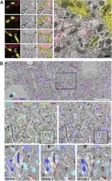
          There is no author summary for this article yet. Authors can add summaries to their articles on ScienceOpen to make them more accessible to a non-specialist audience.

          Abstract

          Array tomography encompasses light and electron microscopy modalities that offer unparalleled opportunities to explore three-dimensional cellular architectures in extremely fine structural and molecular detail. Fluorescence array tomography achieves much higher resolution and molecular multiplexing than most other fluorescence microscopy methods, while electron array tomography can capture three-dimensional ultrastructure much more easily and rapidly than traditional serial-section electron microscopy methods. A correlative fluorescence/electron microscopy mode of array tomography furthermore offers a unique capacity to merge the molecular discrimination strengths of multichannel fluorescence microscopy with the ultrastructural imaging strengths of electron microscopy. This essay samples the first decade of array tomography, highlighting applications in neuroscience.

          Electronic supplementary material

          The online version of this article (10.1186/s12915-018-0560-1) contains supplementary material, which is available to authorized users.

          Related collections

          Most cited references65

          • Record: found
          • Abstract: found
          • Article: not found

          Astrocyte glypicans 4 and 6 promote formation of excitatory synapses via GluA1 AMPA receptors

          In the developing central nervous system (CNS), the control of synapse number and function is critical to the formation of neural circuits. We previously demonstrated that astrocyte-secreted factors powerfully induce the formation of functional excitatory synapses between CNS neurons 1 . Astrocyte-secreted thrombospondins induce structural synapses, however these synapses are post-synaptically silent 2 . Here we use biochemical fractionation of astrocyte conditioned media (ACM) to identify glypican 4 (Gpc4) and 6 (Gpc6) as astrocyte-secreted signals sufficient to induce functional synapses between purified retinal ganglion cell (RGC) neurons, and show that depletion of these molecules from ACM significantly reduces its ability to induce postsynaptic activity. Application of Gpc4 to purified neurons is sufficient to increase the frequency and amplitude of glutamatergic synaptic events. This is achieved by increasing the surface level and clustering, but not overall cellular protein level, of the GluA1 subunit of the AMPA glutamate receptor (AMPAR). Gpc4&6 are expressed by astrocytes in vivo in the developing CNS, with Gpc4 expression enriched in the hippocampus and Gpc6 in the cerebellum. Finally, we demonstrate that Gpc4-deficient mice have defective synapse formation, with decreased amplitude of excitatory synaptic currents in the developing hippocampus and reduced recruitment of AMPARs to synapses. These data identify glypicans as a family of novel astrocyte-derived molecules that are necessary and sufficient to promote glutamate receptor clustering and receptivity and induce the formation of post-synaptically functioning CNS synapses.
            Bookmark
            • Record: found
            • Abstract: found
            • Article: not found

            Protein localization in electron micrographs using fluorescence nanoscopy

            A complete portrait of a cell requires a detailed description of its molecular topography: proteins must be linked to particular organelles. Immuno-electron microscopy can reveal locations of proteins with nanometer resolution but is limited by the quality of fixation, the paucity of antibodies, and the inaccessibility of the antigens. Here, we describe correlative fluorescence electron microscopy for the nanoscopic localization of proteins in electron micrographs. Proteins tagged with Citrine or tdEos were expressed in Caenorhabditis elegans, fixed and embedded. Tagged proteins were imaged from ultrathin sections using stimulated emission depletion microscopy (STED) or photoactivated localization microscopy (PALM). Fluorescence was correlated with organelles imaged in electron micrographs from the same sections. These methods were used to successfully localize histones, a mitochondrial protein, and a presynaptic dense projection protein in electron micrographs.
              Bookmark
              • Record: found
              • Abstract: found
              • Article: not found

              Elastic volume reconstruction from series of ultra-thin microscopy sections.

              Anatomy of large biological specimens is often reconstructed from serially sectioned volumes imaged by high-resolution microscopy. We developed a method to reassemble a continuous volume from such large section series that explicitly minimizes artificial deformation by applying a global elastic constraint. We demonstrate our method on a series of transmission electron microscopy sections covering the entire 558-cell Caenorhabditis elegans embryo and a segment of the Drosophila melanogaster larval ventral nerve cord.
                Bookmark

                Author and article information

                Contributors
                stephens@alleninstitute.org
                Journal
                BMC Biol
                BMC Biol
                BMC Biology
                BioMed Central (London )
                1741-7007
                6 September 2018
                6 September 2018
                2018
                : 16
                : 98
                Affiliations
                GRID grid.417881.3, Allen Institute for Brain Science, ; Seattle, WA USA
                Article
                560
                10.1186/s12915-018-0560-1
                6127925
                30189863
                9a66f4b0-1561-495d-8c3a-7327bddeb1b5
                © Smith et al. 2018

                Open AccessThis article is distributed under the terms of the Creative Commons Attribution 4.0 International License ( http://creativecommons.org/licenses/by/4.0/), which permits unrestricted use, distribution, and reproduction in any medium, provided you give appropriate credit to the original author(s) and the source, provide a link to the Creative Commons license, and indicate if changes were made. The Creative Commons Public Domain Dedication waiver ( http://creativecommons.org/publicdomain/zero/1.0/) applies to the data made available in this article, unless otherwise stated.

                History
                Funding
                Funded by: Allen Institute for Brain Science founders
                Funded by: FundRef http://dx.doi.org/10.13039/100000009, Foundation for the National Institutes of Health;
                Award ID: R01NS092474 and R01MH104227
                Categories
                Question and Answer
                Custom metadata
                © The Author(s) 2018

                Life sciences
                Life sciences

                Comments

                Comment on this article