15
views
0
recommends
+1 Recommend
0 collections
    0
    shares
      • Record: found
      • Abstract: found
      • Article: found
      Is Open Access

      Aldolase A promotes epithelial‐mesenchymal transition to increase malignant potentials of cervical adenocarcinoma

      research-article

      Read this article at

          There is no author summary for this article yet. Authors can add summaries to their articles on ScienceOpen to make them more accessible to a non-specialist audience.

          Abstract

          Recent studies have revealed that metabolic reprogramming is closely associated with epithelial‐mesenchymal transition (EMT) during cancer progression. Aldolase A (ALDOA) is a key glycolytic enzyme that is highly expressed in several types of cancer. In this study, we found that ALDOA is highly expressed in uterine cervical adenocarcinoma and that high ALDOA expression promotes EMT to increase malignant potentials, such as metastasis and invasiveness, in cervical adenocarcinoma cells. In human surgical specimens, ALDOA was highly expressed in cervical adenocarcinoma and high ALDOA expression was correlated with lymph node metastasis, lymphovascular infiltration, and short overall survival. Suppression of ALDOA expression significantly reduced cell growth, migration, and invasiveness of cervical cancer cells. Aldolase A expression was partially regulated by hypoxia‐inducible factor‐1α (HIF‐1α). Shotgun proteome analysis revealed that cell‐cell adhesion‐related proteins were significantly increased in ALDOA‐overexpressing cells. Interestingly, overexpression of ALDOA caused severe morphological changes, including a cuboidal‐to‐spindle shape shift and reduced microvilli formation, coincident with modulation of the expression of typical EMT‐related proteins. Overexpression of ALDOA increased migration and invasion in vitro. Furthermore, overexpression of ALDOA induced HIF‐1α, suggesting a positive feedback loop between ALDOA and HIF‐1α. In conclusion, ALDOA is overexpressed in cervical adenocarcinoma and contributes to malignant potentials of tumor cells through modulation of HIF‐1α signaling. The feedback loop between ALDOA and HIF‐1α could become a therapeutic target to improve the prognosis of this malignancy.

          Abstract

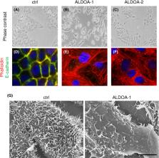
          Aldolase A (ALDOA) overexpression caused epithelial‐mesenchymal transition‐like morphological alterations in cervical adenocarcinoma cells (A‐C). ALDOA‐overexpressing cells showed increased stress fiber formation (D‐F, red) and reduced E‐cadherin expression (D‐F, green) and microvilli formation (G).

          Related collections

          Most cited references43

          • Record: found
          • Abstract: found
          • Article: found
          Is Open Access

          Metabolic reprogramming: the emerging concept and associated therapeutic strategies

          Go Yoshida (2015)
          Tumor tissue is composed of cancer cells and surrounding stromal cells with diverse genetic/epigenetic backgrounds, a situation known as intra-tumoral heterogeneity. Cancer cells are surrounded by a totally different microenvironment than that of normal cells; consequently, tumor cells must exhibit rapidly adaptive responses to hypoxia and hypo-nutrient conditions. This phenomenon of changes of tumor cellular bioenergetics, called “metabolic reprogramming”, has been recognized as one of 10 hallmarks of cancer. Metabolic reprogramming is required for both malignant transformation and tumor development, including invasion and metastasis. Although the Warburg effect has been widely accepted as a common feature of metabolic reprogramming, accumulating evidence has revealed that tumor cells depend on mitochondrial metabolism as well as aerobic glycolysis. Remarkably, cancer-associated fibroblasts in tumor stroma tend to activate both glycolysis and autophagy in contrast to neighboring cancer cells, which leads to a reverse Warburg effect. Heterogeneity of monocarboxylate transporter expression reflects cellular metabolic heterogeneity with respect to the production and uptake of lactate. In tumor tissue, metabolic heterogeneity induces metabolic symbiosis, which is responsible for adaptation to drastic changes in the nutrient microenvironment resulting from chemotherapy. In addition, metabolic heterogeneity is responsible for the failure to induce the same therapeutic effect against cancer cells as a whole. In particular, cancer stem cells exhibit several biological features responsible for resistance to conventional anti-tumor therapies. Consequently, cancer stem cells tend to form minimal residual disease after chemotherapy and exhibit metastatic potential with additional metabolic reprogramming. This type of altered metabolic reprogramming leads to adaptive/acquired resistance to anti-tumor therapy. Collectively, complex and dynamic metabolic reprogramming should be regarded as a reflection of the “robustness” of tumor cells against unfavorable conditions. This review focuses on the concept of metabolic reprogramming in heterogeneous tumor tissue, and further emphasizes the importance of developing novel therapeutic strategies based on drug repositioning.
            Bookmark
            • Record: found
            • Abstract: found
            • Article: not found

            The rising incidence of adenocarcinoma relative to squamous cell carcinoma of the uterine cervix in the United States--a 24-year population-based study.

            The aim of this study was to compare the age-adjusted incidence and survival for invasive adenocarcinoma and squamous cell carcinoma of the uterine cervix using population-based data. The SEER database was used to identify all cases of cervical cancer registered between 1973 and 1996. Stage was defined as localized, regional, or distant. Age-adjusted incidence rates were analyzed statistically using the Jonchkeere-Terpstra exact test for trends. Relative and observed survival rates, respectively, were compared using z tests and log-rank tests. The age-adjusted incidence rates per 100,000 for all invasive cervical cancers decreased by 36.9% over 24 years [12.35 (1973-1977) vs 7.79 (1993-1996)]. Similarly, the age-adjusted incidence rates for squamous cell carcinoma declined by 41.9% [9.45 (1973-1977) vs 5.49 (1993-1996)]. In contrast, the age-adjusted incidence rates for adenocarcinoma increased by 29.1% [1.34 (1973-1977) vs 1.73 (1993-1996)]. The proportion of adenocarcinoma increased 107.4% relative to all cervical cancer, 95.2% relative to squamous cell carcinoma, and 49.3% relative to the population of women at risk [10. 8% vs 22.4% (P < 0.001), 12.4% vs 24.0% (P < 0.001), and 1.40 vs 2. 09 per 100,000 women (P < 0.001), respectively]. Observed survival rates for adenocarcinoma vs squamous cell carcinoma were poorer for regional (P = 0.04), but not localized or distant disease. Over the past 24 years, the incidence of all cervical cancer and squamous cell carcinoma has continued to decline. However, the proportion of adenocarcinoma relative to squamous cell carcinoma and to all cervical cancers has doubled, and the rate of adenocarcinoma per population at risk has also increased. These results suggest that current screening practices in the United States are insufficient to detect a significant proportion of adenocarcinoma precursor lesions. Copyright 2000 Academic Press.
              Bookmark
              • Record: found
              • Abstract: found
              • Article: not found

              Leaving the neighborhood: molecular mechanisms involved during epithelial-mesenchymal transition.

              P Savagner (2001)
              Several molecular mechanisms contribute directly and mechanically to the loss of epithelial phenotype. During epithelial-mesenchymal transition (EMT), adherens junctions and desmosomes are at least partially dissociated. At the same time, a massive cytoskeleton reorganization takes place, involving the rho family and the remodeling of the actin microfilament mesh. Numerous pathways have been described in vitro that control phenotype transition in specific cell models. In vivo developmental studies suggest that transcriptional control, activated by a specific pathway involving Ras, Src and potentially the Wnt pathway, is an essential step. Recent functional and localization experiments indicate that the slug/snail family of transcription factors functions overall as an epithelial phenotype repressor and could represent a key EMT contributor. Copyright 2001 John Wiley & Sons, Inc.
                Bookmark

                Author and article information

                Contributors
                atakasawa@sapmed.ac.jp
                Journal
                Cancer Sci
                Cancer Sci
                10.1111/(ISSN)1349-7006
                CAS
                Cancer Science
                John Wiley and Sons Inc. (Hoboken )
                1347-9032
                1349-7006
                30 June 2020
                August 2020
                : 111
                : 8 ( doiID: 10.1111/cas.v111.8 )
                : 3071-3081
                Affiliations
                [ 1 ] Department of Pathology Sapporo Medical University School of Medicine Sapporo Japan
                [ 2 ] Department of Obstetrics and Gynecology Sapporo Medical University School of Medicine Sapporo Japan
                [ 3 ] Department of Surgical Pathology Sapporo Medical University School of Medicine Sapporo Japan
                Author notes
                [*] [* ] Correspondence

                Akira Takasawa, Department of Pathology, Sapporo Medical University School of Medicine, Sapporo, Japan.

                Email: atakasawa@ 123456sapmed.ac.jp

                Author information
                https://orcid.org/0000-0002-3244-1199
                https://orcid.org/0000-0002-0608-3914
                https://orcid.org/0000-0003-1754-252X
                Article
                CAS14524
                10.1111/cas.14524
                7419050
                32530543
                8b8233e5-2e16-462f-a5f7-3b4f75f0bbc1
                © 2020 The Authors. Cancer Science published by John Wiley & Sons Australia, Ltd on behalf of Japanese Cancer Association.

                This is an open access article under the terms of the http://creativecommons.org/licenses/by-nc-nd/4.0/ License, which permits use and distribution in any medium, provided the original work is properly cited, the use is non‐commercial and no modifications or adaptations are made.

                History
                : 27 December 2019
                : 30 May 2020
                : 08 June 2020
                Page count
                Figures: 6, Tables: 1, Pages: 11, Words: 6475
                Funding
                Funded by: The Suhara Foundation
                Funded by: Japan Society for the Promotion of Science , open-funder-registry 10.13039/501100001691;
                Award ID: JP17K08697
                Award ID: JP17K08698
                Award ID: JP18K15084
                Award ID: JP19K16561
                Categories
                Original Article
                Original Articles
                Pathology
                Custom metadata
                2.0
                August 2020
                Converter:WILEY_ML3GV2_TO_JATSPMC version:5.8.6 mode:remove_FC converted:11.08.2020

                Oncology & Radiotherapy
                aldolase a,cervical adenocarcinoma,epithelial‐mesenchymal transition,hypoxia‐inducible factor‐1α,metabolic reprogramming

                Comments

                Comment on this article