4
views
0
recommends
+1 Recommend
0 collections
    0
    shares
      • Record: found
      • Abstract: found
      • Article: found
      Is Open Access

      The Long Noncoding RNA Hotair Regulates Oxidative Stress and Cardiac Myocyte Apoptosis during Ischemia-Reperfusion Injury

      research-article

      Read this article at

          There is no author summary for this article yet. Authors can add summaries to their articles on ScienceOpen to make them more accessible to a non-specialist audience.

          Abstract

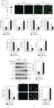
          Oxidative stress and subsequent cardiac myocyte apoptosis play central roles in the initiation and progression of myocardial ischemia-reperfusion (I/R) injury. Homeobox transcript antisense intergenic RNA ( Hotair) was previously implicated in various heart diseases, yet its role in myocardial I/R injury has not been clearly demonstrated. Mice with cardiac-restricted knockdown or overexpression of Hotair were exposed to I/R surgery. H9c2 cells were cultured and subjected to hypoxia/reoxygenation (H/R) stimulation to further verify the role and underlying mechanisms of Hotair in vitro. Histological examination, molecular detection, and functional parameters were determined in vivo and in vitro. In response to I/R or H/R treatment, Hotair expression was increased in a bromodomain-containing protein 4-dependent manner. Cardiac-restricted knockdown of Hotair exacerbated, whereas Hotair overexpression prevented I/R-induced oxidative stress, cardiac myocyte apoptosis, and cardiac dysfunction. Mechanistically, we observed that Hotair exerted its beneficial effects via activating AMP-activated protein kinase alpha (AMPK α). Further detection revealed that Hotair activated AMPK α through regulating the enhancer of zeste homolog 2/microRNA-451/calcium-binding protein 39 (EZH2/ miR-451/Cab39) axis. We provide the evidence that endogenous lncRNA Hotair is an essential negative regulator for oxidative stress and cardiac myocyte apoptosis in myocardial I/R injury, which is dependent on AMPK α activation via the EZH2/ miR-451/Cab39 axis.

          Related collections

          Most cited references42

          • Record: found
          • Abstract: found
          • Article: found
          Is Open Access

          Pathogenesis of cardiac ischemia reperfusion injury is associated with CK2α-disturbed mitochondrial homeostasis via suppression of FUNDC1-related mitophagy

          Disturbed mitochondrial homeostasis contributes to the pathogenesis of cardiac ischemia reperfusion (IR) injury, although the underlying mechanism remains elusive. Here, we demonstrated that casein kinase 2α (CK2α) was upregulated following acute cardiac IR injury. Increased CK2α was shown to be instrumental to mitochondrial damage, cardiomyocyte death, infarction area expansion and cardiac dysfunction, whereas cardiac-specific CK2α knockout (CK2α CKO ) mice were protected against IR injury and mitochondrial damage. Functional assay indicated that CK2α enhanced the phosphorylation (inactivation) of FUN14 domain containing 1 (FUNDC1) via post-transcriptional modification at Ser13, thus effectively inhibiting mitophagy. Defective mitophagy failed to remove damaged mitochondria induced by IR injury, resulting in mitochondrial genome collapse, electron transport chain complex (ETC) inhibition, mitochondrial biogenesis arrest, cardiolipin oxidation, oxidative stress, mPTP opening, mitochondrial debris accumulation and eventually mitochondrial apoptosis. In contrast, loss of CK2α reversed the FUNDC1-mediated mitophagy, providing a survival advantage to myocardial tissue following IR stress. Interestingly, mice deficient in both CK2α and FUNDC1 failed to show protection against IR injury and mitochondrial damage through a mechanism possible attributed to lack of mitophagy. Taken together, our results confirmed that CK2α serves as a negative regulator of mitochondrial homeostasis via suppression of FUNDC1-required mitophagy, favoring the development of cardiac IR injury.
            Bookmark
            • Record: found
            • Abstract: not found
            • Article: not found

            Melatonin attenuates myocardial ischemia‐reperfusion injury via improving mitochondrial fusion/mitophagy and activating the AMPK‐OPA1 signaling pathways

              Bookmark
              • Record: found
              • Abstract: found
              • Article: not found

              AMP-activated protein kinase mediates ischemic glucose uptake and prevents postischemic cardiac dysfunction, apoptosis, and injury.

              AMP-activated protein kinase (AMPK) is an important regulator of diverse cellular pathways in the setting of energetic stress. Whether AMPK plays a critical role in the metabolic and functional responses to myocardial ischemia and reperfusion remains uncertain. We examined the cardiac consequences of long-term inhibition of AMPK activity in transgenic mice expressing a kinase dead (KD) form of the enzyme. The KD mice had normal fractional shortening and no heart failure, cardiac hypertrophy, or fibrosis, although the in vivo left ventricular (LV) dP/dt was lower than that in WT hearts. During low-flow ischemia and postischemic reperfusion in vitro, KD hearts failed to augment glucose uptake and glycolysis, although glucose transporter content and insulin-stimulated glucose uptake were normal. KD hearts also failed to increase fatty acid oxidation during reperfusion. Furthermore, KD hearts demonstrated significantly impaired recovery of LV contractile function during postischemic reperfusion that was associated with a lower ATP content and increased injury compared with WT hearts. Caspase-3 activity and TUNEL-staining were increased in KD hearts after ischemia and reperfusion. Thus, AMPK is responsible for activation of glucose uptake and glycolysis during low-flow ischemia and plays an important protective role in limiting damage and apoptotic activity associated with ischemia and reperfusion in the heart.
                Bookmark

                Author and article information

                Contributors
                Journal
                Oxid Med Cell Longev
                Oxid Med Cell Longev
                OMCL
                Oxidative Medicine and Cellular Longevity
                Hindawi
                1942-0900
                1942-0994
                2020
                12 March 2020
                : 2020
                : 1645249
                Affiliations
                Department of Cardiology, Union Hospital, Tongji Medical College, Huazhong University of Science and Technology, Wuhan 430022, China
                Author notes

                Guest Editor: Tatjana Bačun

                Author information
                https://orcid.org/0000-0002-6472-8450
                https://orcid.org/0000-0001-6081-3327
                https://orcid.org/0000-0003-3713-9930
                https://orcid.org/0000-0003-4867-3805
                https://orcid.org/0000-0002-6791-6545
                Article
                10.1155/2020/1645249
                7091551
                9e820da8-fa55-42af-8f44-5ce90354ff43
                Copyright © 2020 Kai Meng et al.

                This is an open access article distributed under the Creative Commons Attribution License, which permits unrestricted use, distribution, and reproduction in any medium, provided the original work is properly cited.

                History
                : 16 December 2019
                : 3 February 2020
                : 17 February 2020
                Funding
                Funded by: National Natural Science Foundation of China
                Award ID: 81800439
                Award ID: 81600231
                Categories
                Research Article

                Molecular medicine
                Molecular medicine

                Comments

                Comment on this article