- Record: found
- Abstract: found
- Article: not found
Altered decorin expression of systemic sclerosis by UVA1 (340–400 nm) phototherapy: Immunohistochemical analysis of 3 cases
Read this article at

Abstract
Background
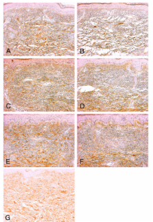
Ultraviolet A1 (340–400 nm, UVA1) phototherapy is highly effective in sclerotic lesions of systemic sclerosis (SSc). Histological evaluation of skin specimens obtained before and after UVA1 phototherapy revealed loosening of collagen bundles and the appearance of small collagen fibers. We have previously shown that UVA1 irradiation induced collagenase in vitro study by using SSc fibroblasts. The increased levels of mRNA and protein of decorin in SSc fibroblasts were reported. In this study, we focus on the lesional expression of small dermatan sulfate proteoglycan, decorin that has a role of binding to collagen and fibrillogenesis.
Case presentation
We employed immunohistochemical analysis of decorin before and after UVA1 phototherapy. The skin specimens from three patients who were effectively treated with UVA1 phototherapy were analysed. Monoclonal antibody 6B6 as the specific reactivity to decorin was used. The increased decorin was focally accumulated in the newly synthesized collagen fibers in the sclerotic lesion of SSc. After UVA1 phototherapy, decorin was decreased in upper to middle dermis, although decorin was slightly increased in papillary dermis.
Related collections
Most cited references19
- Record: found
- Abstract: found
- Article: not found
Natural inhibitor of transforming growth factor-beta protects against scarring in experimental kidney disease.
- Record: found
- Abstract: not found
- Article: not found
The biology of the small leucine-rich proteoglycans. Functional network of interactive proteins.
- Record: found
- Abstract: found
- Article: not found