66
views
0
recommends
+1 Recommend
0 collections
    0
    shares
      • Record: found
      • Abstract: found
      • Article: found
      Is Open Access

      Sustained production of ROS triggers compensatory proliferation and is required for regeneration to proceed

      research-article

      Read this article at

          There is no author summary for this article yet. Authors can add summaries to their articles on ScienceOpen to make them more accessible to a non-specialist audience.

          Abstract

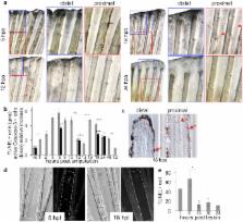
          A major issue in regenerative medicine is the role of injury in promoting cell plasticity. Here we explore the function of reactive oxygen species (ROS) induced through lesions in adult zebrafish. We show that ROS production, following adult fin amputation, is tightly regulated in time and space for at least 24 hours, whereas ROS production remains transient (2 hours) in mere wound healing. In regenerative tissue, ROS signaling triggers two distinct parallel pathways: one pathway is responsible for apoptosis, and the other pathway is responsible for JNK activation. Both events are involved in the compensatory proliferation of stump epidermal cells and are necessary for the progression of regeneration. Both events impact the Wnt, SDF1 and IGF pathways, while apoptosis only impacts progenitor marker expression. These results implicate oxidative stress in regeneration and provide new insights into the differences between healing and regeneration.

          Related collections

          Most cited references45

          • Record: found
          • Abstract: found
          • Article: not found

          A tissue-scale gradient of hydrogen peroxide mediates rapid wound detection in zebrafish

          Barrier structures (e.g. epithelia around tissues, plasma membranes around cells) are required for internal homeostasis and protection from pathogens. Wound detection and healing represent a dormant morphogenetic program that can be rapidly executed to restore barrier integrity and tissue homeostasis. In animals, initial steps include recruitment of leukocytes to the site of injury across distances of hundreds of micrometers within minutes of wounding. The spatial signals that direct this immediate tissue response are unknown. Due to their fast diffusion and versatile biological activities, reactive oxygen species (ROS), including hydrogen peroxide (H2O2), are interesting candidates for wound-to-leukocyte signalling. We probed the role of H2O2 during the early events of wound responses in zebrafish larvae expressing a genetically encoded H2O2 sensor1. This reporter revealed a sustained rise in H2O2 concentration at the wound margin, starting ∼3 min after wounding and peaking at ∼20 min, which extended ∼100−200 μm into the tail fin epithelium as a decreasing concentration gradient. Using pharmacological and genetic inhibition, we show that this gradient is created by Dual oxidase (Duox), and that it is required for rapid recruitment of leukocytes to the wound. This is the first observation of a tissue-scale H2O2 pattern, and the first evidence that H2O2 signals to leukocytes in tissues, in addition to its known antiseptic role.
            Bookmark
            • Record: found
            • Abstract: found
            • Article: not found

            Reactive oxygen species promote TNFalpha-induced death and sustained JNK activation by inhibiting MAP kinase phosphatases.

            TNFalpha is a pleiotropic cytokine that induces either cell proliferation or cell death. Inhibition of NF-kappaB activation increases susceptibility to TNFalpha-induced death, concurrent with sustained JNK activation, an important contributor to the death response. Sustained JNK activation in NF-kappaB-deficient cells was suggested to depend on reactive oxygen species (ROS), but how ROS affect JNK activation was unclear. We now show that TNFalpha-induced ROS, whose accumulation is suppressed by mitochondrial superoxide dismutase, cause oxidation and inhibition of JNK-inactivating phosphatases by converting their catalytic cysteine to sulfenic acid. This results in sustained JNK activation, which is required for cytochrome c release and caspase 3 cleavage, as well as necrotic cell death. Treatment of cells or experimental animals with an antioxidant prevents H(2)O(2) accumulation, JNK phosphatase oxidation, sustained JNK activity, and both forms of cell death. Antioxidant treatment also prevents TNFalpha-mediated fulminant liver failure without affecting liver regeneration.
              Bookmark
              • Record: found
              • Abstract: found
              • Article: not found

              Cells keep a memory of their tissue origin during axolotl limb regeneration.

              During limb regeneration adult tissue is converted into a zone of undifferentiated progenitors called the blastema that reforms the diverse tissues of the limb. Previous experiments have led to wide acceptance that limb tissues dedifferentiate to form pluripotent cells. Here we have reexamined this question using an integrated GFP transgene to track the major limb tissues during limb regeneration in the salamander Ambystoma mexicanum (the axolotl). Surprisingly, we find that each tissue produces progenitor cells with restricted potential. Therefore, the blastema is a heterogeneous collection of restricted progenitor cells. On the basis of these findings, we further demonstrate that positional identity is a cell-type-specific property of blastema cells, in which cartilage-derived blastema cells harbour positional identity but Schwann-derived cells do not. Our results show that the complex phenomenon of limb regeneration can be achieved without complete dedifferentiation to a pluripotent state, a conclusion with important implications for regenerative medicine.
                Bookmark

                Author and article information

                Journal
                Sci Rep
                Sci Rep
                Scientific Reports
                Nature Publishing Group
                2045-2322
                27 June 2013
                2013
                : 3
                : 2084
                Affiliations
                [1 ]Université Paris Diderot , Sorbonne Paris Cité. Paris - France
                [2 ]Centre Interdisciplinaire de Recherche en biologie (CIRB) CNRS UMR 7241/INSERM U1050/Collège de France. Paris - France
                [3 ]Department of Biology - Ecole Normale Supérieure. Paris – France
                [4 ]These authors contributed equally to this work.
                [5 ]Current address: Department of Molecular, Cellular, and Developmental Biology Yale University- New Haven, CT, USA.
                Author notes
                Article
                srep02084
                10.1038/srep02084
                3694286
                23803955
                b593890a-205c-4047-b9b5-4c7d757ede68
                Copyright © 2013, Macmillan Publishers Limited. All rights reserved

                This work is licensed under a Creative Commons Attribution-NonCommercial-NoDerivs 3.0 Unported License. To view a copy of this license, visit http://creativecommons.org/licenses/by-nc-nd/3.0/

                History
                : 06 March 2013
                : 10 June 2013
                Categories
                Article

                Uncategorized
                Uncategorized

                Comments

                Comment on this article