25
views
0
recommends
+1 Recommend
0 collections
    0
    shares
      • Record: found
      • Abstract: found
      • Article: found
      Is Open Access

      Structural disconnection and functional reorganization in Fabry disease: a multimodal MRI study

      research-article

      Read this article at

          There is no author summary for this article yet. Authors can add summaries to their articles on ScienceOpen to make them more accessible to a non-specialist audience.

          Abstract

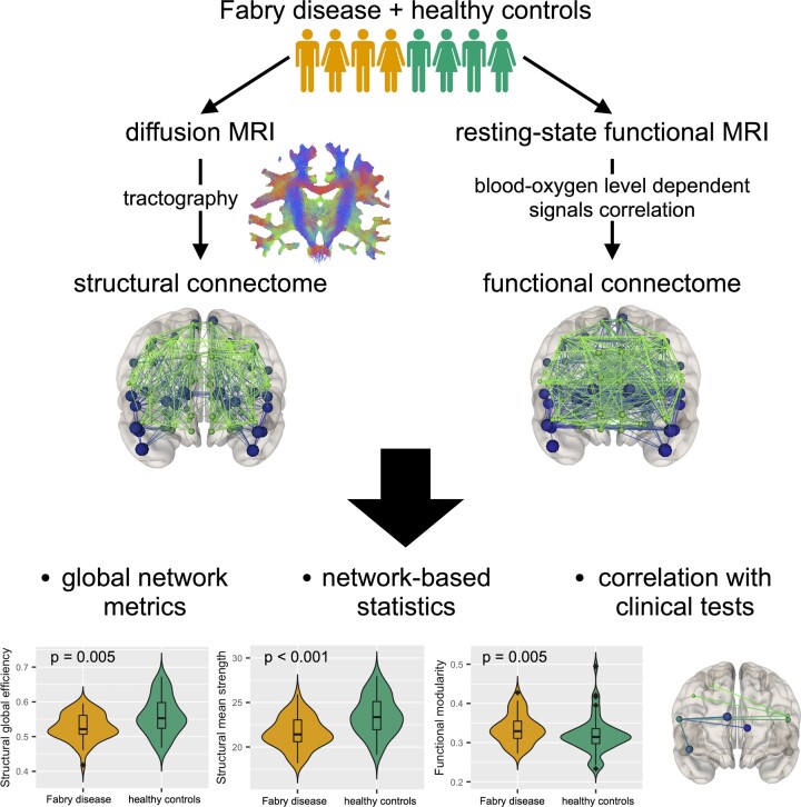
          Central nervous system involvement in Fabry disease, a rare systemic X-linked lysosomal storage disorder, is characterized by the presence of heterogeneous but consistent functional and microstructural changes. Nevertheless, knowledge about the degree and extension of macro-scale brain connectivity modifications is to date missing. In this work, we performed connectomic analyses of diffusion and resting-state functional MRI to investigate changes of both structural and functional brain organization in Fabry disease, as well as to explore the relationship between the two and their clinical correlates. In this retrospective cross-sectional study, 46 patients with Fabry disease (28F, 42.2 ± 13.2years) and 49 healthy controls (21F, 42.3 ± 16.3years) were included. All subjects underwent an MRI examination including anatomical, diffusion and resting-state functional sequences. Images were processed to obtain quantitative structural and functional connectomes, where the connections between regions of interest were weighted by the total intra-axonal signal contribution of the corresponding bundle and by the correlation between blood-oxygen level–dependent time series, respectively. We explored between-group differences in terms of both global network properties, expressed with graph measures and specific connected subnetworks, identified using a network-based statistics approach. As exploratory analyses, we also investigated the possible association between cognitive performance and structural and functional connectome modifications at both global and subnetwork level in a subgroup of patients ( n = 11). Compared with healthy controls, patients with Fabry disease showed a significantly reduced global efficiency ( P = 0.005) and mean strength ( P < 0.001) in structural connectomes, together with an increased modularity ( P = 0.005) in functional networks. As for the network-based statistics analysis, a subnetwork with decreased structural connectivity in patients with Fabry disease compared with healthy controls emerged, with eight nodes mainly located at the level of frontal or deep grey-matter areas. When probing the relation between altered global network metrics and neuropsychological tests, correlations emerged between the structural and functional disruption with results at verbal and working memory tests, respectively. Furthermore, structural disruption at subnetwork level was associated with worse executive functioning, with a significant moderation effect of functional changes suggesting a compensation mechanism. Taken together, these results further expand the current knowledge about brain involvement in Fabry disease, showing widespread structural disconnection and functional reorganization, primarily sustained by loss in axonal integrity and correlating with cognitive performance.

          Abstract

          Gabusi et al. examined structural and functional changes of brain connectivity in Fabry disease using diffusion and resting-state functional MRI data. The connectome analysis of brain networks revealed that patients with Fabry disease show both a structural disconnection (due to mild but widespread axonal damage) and a functional reorganization, associated to cognitive performances.

          Graphical Abstract

          Graphical abstract

          Related collections

          Most cited references70

          • Record: found
          • Abstract: found
          • Article: not found

          Automated anatomical labeling of activations in SPM using a macroscopic anatomical parcellation of the MNI MRI single-subject brain.

          An anatomical parcellation of the spatially normalized single-subject high-resolution T1 volume provided by the Montreal Neurological Institute (MNI) (D. L. Collins et al., 1998, Trans. Med. Imag. 17, 463-468) was performed. The MNI single-subject main sulci were first delineated and further used as landmarks for the 3D definition of 45 anatomical volumes of interest (AVOI) in each hemisphere. This procedure was performed using a dedicated software which allowed a 3D following of the sulci course on the edited brain. Regions of interest were then drawn manually with the same software every 2 mm on the axial slices of the high-resolution MNI single subject. The 90 AVOI were reconstructed and assigned a label. Using this parcellation method, three procedures to perform the automated anatomical labeling of functional studies are proposed: (1) labeling of an extremum defined by a set of coordinates, (2) percentage of voxels belonging to each of the AVOI intersected by a sphere centered by a set of coordinates, and (3) percentage of voxels belonging to each of the AVOI intersected by an activated cluster. An interface with the Statistical Parametric Mapping package (SPM, J. Ashburner and K. J. Friston, 1999, Hum. Brain Mapp. 7, 254-266) is provided as a freeware to researchers of the neuroimaging community. We believe that this tool is an improvement for the macroscopical labeling of activated area compared to labeling assessed using the Talairach atlas brain in which deformations are well known. However, this tool does not alleviate the need for more sophisticated labeling strategies based on anatomical or cytoarchitectonic probabilistic maps.
            Bookmark
            • Record: found
            • Abstract: found
            • Article: not found

            The organization of the human cerebral cortex estimated by intrinsic functional connectivity.

            Information processing in the cerebral cortex involves interactions among distributed areas. Anatomical connectivity suggests that certain areas form local hierarchical relations such as within the visual system. Other connectivity patterns, particularly among association areas, suggest the presence of large-scale circuits without clear hierarchical relations. In this study the organization of networks in the human cerebrum was explored using resting-state functional connectivity MRI. Data from 1,000 subjects were registered using surface-based alignment. A clustering approach was employed to identify and replicate networks of functionally coupled regions across the cerebral cortex. The results revealed local networks confined to sensory and motor cortices as well as distributed networks of association regions. Within the sensory and motor cortices, functional connectivity followed topographic representations across adjacent areas. In association cortex, the connectivity patterns often showed abrupt transitions between network boundaries. Focused analyses were performed to better understand properties of network connectivity. A canonical sensory-motor pathway involving primary visual area, putative middle temporal area complex (MT+), lateral intraparietal area, and frontal eye field was analyzed to explore how interactions might arise within and between networks. Results showed that adjacent regions of the MT+ complex demonstrate differential connectivity consistent with a hierarchical pathway that spans networks. The functional connectivity of parietal and prefrontal association cortices was next explored. Distinct connectivity profiles of neighboring regions suggest they participate in distributed networks that, while showing evidence for interactions, are embedded within largely parallel, interdigitated circuits. We conclude by discussing the organization of these large-scale cerebral networks in relation to monkey anatomy and their potential evolutionary expansion in humans to support cognition.
              Bookmark
              • Record: found
              • Abstract: found
              • Article: not found

              Complex network measures of brain connectivity: uses and interpretations.

              Brain connectivity datasets comprise networks of brain regions connected by anatomical tracts or by functional associations. Complex network analysis-a new multidisciplinary approach to the study of complex systems-aims to characterize these brain networks with a small number of neurobiologically meaningful and easily computable measures. In this article, we discuss construction of brain networks from connectivity data and describe the most commonly used network measures of structural and functional connectivity. We describe measures that variously detect functional integration and segregation, quantify centrality of individual brain regions or pathways, characterize patterns of local anatomical circuitry, and test resilience of networks to insult. We discuss the issues surrounding comparison of structural and functional network connectivity, as well as comparison of networks across subjects. Finally, we describe a Matlab toolbox (http://www.brain-connectivity-toolbox.net) accompanying this article and containing a collection of complex network measures and large-scale neuroanatomical connectivity datasets. Copyright (c) 2009 Elsevier Inc. All rights reserved.
                Bookmark

                Author and article information

                Contributors
                Journal
                Brain Commun
                Brain Commun
                braincomms
                Brain Communications
                Oxford University Press
                2632-1297
                2022
                22 July 2022
                22 July 2022
                : 4
                : 4
                : fcac187
                Affiliations
                Department of Computer Science, Diffusion Imaging and Connectivity Estimation (DICE) Lab, University of Verona , Verona 37134, Italy
                Department of Advanced Biomedical Sciences, University “Federico II” , Naples 80131, Italy
                Department of Advanced Biomedical Sciences, University “Federico II” , Naples 80131, Italy
                Department of Electrical Engineering and Information Technology (DIETI), University “Federico II” , Naples 80125, Italy
                Department of Human Neuroscience, Sapienza University of Rome , Rome 00189, Italy
                Department of Computer Science, Diffusion Imaging and Connectivity Estimation (DICE) Lab, University of Verona , Verona 37134, Italy
                Department of Computer Science, University of Sherbrooke , Sherbrooke, QC J1K 2R1, Canada
                Department of Computer Science, Diffusion Imaging and Connectivity Estimation (DICE) Lab, University of Verona , Verona 37134, Italy
                Department of Biomedical Engineering, Translational Imaging in Neurology (ThINk), University Hospital Basel and University of Basel , Basel 4001, Switzerland
                Department of Clinical and Experimental Medicine, Multiple Sclerosis Centre, II Division of Neurology, ‘'Luigi Vanvitelli” University , Naples 80138, Italy
                Department of Computer Science, Diffusion Imaging and Connectivity Estimation (DICE) Lab, University of Verona , Verona 37134, Italy
                Department of Neurosciences and Reproductive and Odontostomatological Sciences, University “Federico II” , Naples 80131, Italy
                Department of Public Health, Nephrology Unit, University “Federico II” , Naples 80131, Italy
                Department of Neurosciences and Reproductive and Odontostomatological Sciences, University “Federico II” , Naples 80131, Italy
                Department of Advanced Biomedical Sciences, University “Federico II” , Naples 80131, Italy
                Department of Computer Science, Diffusion Imaging and Connectivity Estimation (DICE) Lab, University of Verona , Verona 37134, Italy
                Department of Neuroscience, Rehabilitation, Ophthalmology, Genetics, Maternal and Child Health (DINOGMI), University of Genoa , Genoa 16132, Italy
                Department of Advanced Biomedical Sciences, University “Federico II” , Naples 80131, Italy
                Author notes
                Correspondence to: Giuseppe Pontillo, MD Department of Advanced Biomedical Sciences Department of Electrical Engineering and Information Technology (DIETI) University ‘Federico II’, Via Pansini 5, 80131 Naples, Italy E-mail: giuseppe.pontillo@ 123456unina.it

                Ilaria Gabusi, Giuseppe Pontillo, Simona Schiavi and Sirio Cocozza contributed equally to this work.

                Author information
                https://orcid.org/0000-0002-2553-8304
                https://orcid.org/0000-0001-5425-1890
                https://orcid.org/0000-0001-9429-2769
                https://orcid.org/0000-0003-1641-186X
                https://orcid.org/0000-0002-0300-5160
                Article
                fcac187
                10.1093/braincomms/fcac187
                9327118
                ab70aecd-ef74-4e64-8b36-5e470e7f8ff6
                © The Author(s) 2022. Published by Oxford University Press on behalf of the Guarantors of Brain.

                This is an Open Access article distributed under the terms of the Creative Commons Attribution License ( https://creativecommons.org/licenses/by/4.0/), which permits unrestricted reuse, distribution, and reproduction in any medium, provided the original work is properly cited.

                History
                : 03 January 2022
                : 17 May 2022
                : 20 July 2022
                Page count
                Pages: 12
                Categories
                Original Article
                AcademicSubjects/MED00310
                AcademicSubjects/SCI01870

                fabry disease,magnetic resonance imaging,brain connectivity,multimodal study,microstructure informed tractography

                Comments

                Comment on this article