10
views
0
recommends
+1 Recommend
0 collections
    0
    shares
      • Record: found
      • Abstract: found
      • Article: found
      Is Open Access

      Let-7a-5p may participate in the pathogenesis of diabetic nephropathy through targeting HMGA2

      research-article

      Read this article at

          There is no author summary for this article yet. Authors can add summaries to their articles on ScienceOpen to make them more accessible to a non-specialist audience.

          Abstract

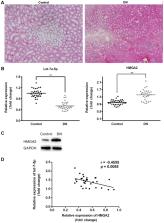
          Diabetic nephropathy (DN) is one of the most common complications of diabetes mellitus (DM), and has been demonstrated as one of the major causes of renal failure. In a previous study, it was noted that microRNA let-7a-5p was downregulated in DN; however, the underlying mechanism requires additional investigation. The aim of the present study was to investigate the roles of let-7a-5p in the pathogenesis of DN and its associated mechanism. The renal tissues of db/db and db/m mice, and renal mesangial cells treated with high concentrations of glucose were obtained; reverse transcription-quantitative polymerase chain reaction, and western blot analysis were applied to detect the expression of let-7a-5p and high-mobility group AT-hook 2 (HMGA2) in vivo and in vitro. In addition, renal mesangial cells cultured under high-glucose conditions (20 and 30 mmol/l) were transfected with either let-7a-5p mimics or let-7a-5p inhibitors. The effects of let-7a-5p on the proliferation and apoptosis of renal mesangial cells were examined using CCK-8 and flow cytometry methods. Additionally, cells were collected and the expression of phosphoinositide 3-kinase (PI3K), phosphorylated protein kinase B (p-AKT) and HMGA2 was analyzed with western blot analysis. Finally, a dual luciferase reporter assay was performed to confirm whether HMGA2 was a direct target of let-7a-5p. Let-7a-5p was significantly downregulated and HMGA2 was markedly upregulated in the tissue samples of DN mice and renal mesangial cells cultured under high-glucose conditions. In addition, transfection of let-7a-5p mimics induced a significant decrease in the proliferation and increase in the apoptosis of renal mesangial cells cultured under high-glucose conditions; transfection of let-7a-5p inhibitors exhibited the opposite effects. Furthermore, transfection of let-7a-5p mimics also led to the inhibition of the PI3K-AKT signaling pathway; transfection of let-7a-5p inhibitors may activate the PI3K-AKT signaling pathway through the increase in PI3K and AKT levels. Finally, a dual luciferase reporter assay confirmed that HMGA2 is a direct target of let-7a-5p. Let-7a-5p was downregulated in DN and may participate in the pathogenesis of DN via regulating HMGA2 expression and the PI3K-AKT signaling pathway.

          Related collections

          Most cited references32

          • Record: found
          • Abstract: found
          • Article: not found

          Diabetic nephropathy: Is there a role for oxidative stress?

          Oxidative stress has been implicated in the pathophysiology of diabetic nephropathy. Studies in experimental animal models of diabetes strongly implicate oxidant species as a major determinant in the pathophysiology of diabetic kidney disease. The translation, in the clinical setting, of these concepts have been quite disappointing, and new theories have challenged the concepts that oxidative stress per se plays a role in the pathophysiology of diabetic kidney disease. The concept of mitochondrial hormesis has been introduced to explain this apparent disconnect. Hormesis is intended as any cellular process that exhibits a biphasic response to exposure to increasing amounts of a substance or condition: specifically, in diabetic kidney disease, oxidant species may represent, at determined concentration, an essential and potentially protective factor. It could be postulated that excessive production or inhibition of oxidant species formation might result in an adverse phenotype. This review discusses the evidence underlying these two apparent contradicting concepts, with the aim to propose and speculate on potential mechanisms underlying the role of oxidant species in the pathophysiology of diabetic nephropathy and possibly open future more efficient therapies to be tested in the clinical settings.
            Bookmark
            • Record: found
            • Abstract: found
            • Article: found
            Is Open Access

            MicroRNA-27a promotes podocyte injury via PPARγ-mediated β-catenin activation in diabetic nephropathy

            Podocyte injury has a pivotal role in the pathogenesis of diabetic nephropathy (DN). MicroRNA-27a (miR-27a), peroxisome proliferator-activated receptor γ (PPARγ) and β-catenin pathways have been involved in the pathogenesis of DN. Herein, we asked whether miR-27a mediates podocyte injury through PPARγ/β-catenin signaling in DN. The functional relevance of miR-27a, PPARγ and β-catenin were investigated in cultured podocytes and glomeruli of diabetic rats and patients using in vitro and in vivo approaches. Podocyte injury was assessed by migration, invasion and apoptosis assay. Biological parameters were analyzed using enzyme-linked immunosorbent assay. We found that high glucose stimulated miR-27a expression, which, by negatively targeting PPARγ, activated β-catenin signaling as evidenced by upregulation of β-catenin target genes, snail1 and α-smooth muscle actin (α-SMA) and downregulation of podocyte-specific markers podocin and synaptopodin. These changes caused podocyte injury as demonstrated by increased podocyte mesenchymal transition, disrupted podocyte architectural integrity and increased podocyte apoptosis. Furthermore, we provide evidence that miR-27a contributed to unfavorable renal function and increased podocyte injury in diabetic rats. Notably, miR-27a exhibited clinical and biological relevance as it was linked to elevated serum creatinine, proteinuria and reduced creatinine clearance rate. In addition, miR-27a upregulation and activation of PPARγ/β-catenin signaling were verified in renal biopsy samples from DN patients. We propose a novel role of the miR-27a/PPARγ/β-catenin axis in fostering the progression toward more deteriorated podocyte injury in DN. Targeting miR-27a could be a potential therapeutic approach for DN.
              Bookmark
              • Record: found
              • Abstract: found
              • Article: found
              Is Open Access

              MiRNA-29c regulates the expression of inflammatory cytokines in diabetic nephropathy by targeting tristetraprolin

              Diabetic nephropathy is one of the most prevalent chronic complications of Diabetes mellitus, but its pathogenesis remains elusive. This study was designed to determine the role of tristetraprolin (TTP), inflammatory cytokines and microRNAs (miRNAs) in DN. The blood and urine samples were obtained from 32 patients with DN, 33 patients with type 2 DM, and 35 normal healthy subjects as controls. Renal tissue samples were also obtained from 10 DN patients and 10 normal controls. The miRNA microarray analyses were performed in pooled plasma and urine sediment samples of eight DN patients and eight age- and sex-matched health control subjects and three paired renal tissues from patients with DN and normal controls. Conditionally immortalized mouse podocytes (MPC5) were used a cell model. The expressions of TTP and cytokines in patient samples and cultured cells were determined by qRT-PCR and Western blotting or ELISA. Our results indicated that miRNA-29c directly targeted TTP and promoted inflammatory response under hyperglycemic conditions. Overexpression of miRNA-29c in podocytes resulted in an increase in inflammatory cytokines and inhibition of miRNA-29c by using its inhibitor reduced the inflammatory cytokines in podocytes. Finally, miRNA-29c promoted the progression of DN by targeting TTP, providing a target for a therapeutic intervention of DN.
                Bookmark

                Author and article information

                Journal
                Mol Med Rep
                Mol Med Rep
                Molecular Medicine Reports
                D.A. Spandidos
                1791-2997
                1791-3004
                May 2019
                19 March 2019
                19 March 2019
                : 19
                : 5
                : 4229-4237
                Affiliations
                [1 ]Department of Clinical Laboratory, Taixing City Second People's Hospital, Taixing, Jiangsu 225400, P.R. China
                [2 ]Department of Endocrinology, Taizhou People's Hospital, Taizhou, Jiangsu 225300, P.R. China
                Author notes
                Correspondence to: Dr Xiaoqiang Fei, Department of Endocrinology, Taizhou People's Hospital, 399 Hailing Road, Taizhou, Jiangsu 225300, P.R. China, E-mail: feixiaoqiang0356@ 123456outlook.com
                Article
                mmr-19-05-4229
                10.3892/mmr.2019.10057
                6471493
                30896854
                d665272c-1e27-43d9-a78d-6b0f7a8aeac2
                Copyright: © Wang et al.

                This is an open access article distributed under the terms of the Creative Commons Attribution-NonCommercial-NoDerivs License, which permits use and distribution in any medium, provided the original work is properly cited, the use is non-commercial and no modifications or adaptations are made.

                History
                : 17 May 2018
                : 26 October 2018
                Categories
                Articles

                microrna,let-7a-5p,diabetic nephropathy,proliferation,apoptosis,high-mobility group at-hook 2

                Comments

                Comment on this article