3
views
0
recommends
+1 Recommend
0 collections
    0
    shares
      • Record: found
      • Abstract: found
      • Article: found
      Is Open Access

      Molecular and cytological profiling of biological aging of mouse cochlear inner and outer hair cells

      research-article

      Read this article at

          There is no author summary for this article yet. Authors can add summaries to their articles on ScienceOpen to make them more accessible to a non-specialist audience.

          SUMMARY

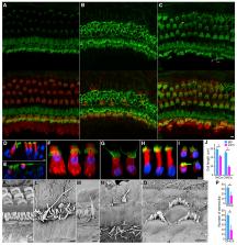
          Age-related hearing loss (ARHL) negatively impacts quality of life in the elderly population. The prevalent cause of ARHL is loss of mechanosensitive cochlear hair cells (HCs). The molecular and cellular mechanisms of HC degeneration remain poorly understood. Using RNA-seq transcriptomic analyses of inner and outer HCs isolated from young and aged mice, we show that HC aging is associated with changes in key molecular processes, including transcription, DNA damage, autophagy, and oxidative stress, as well as genes related to HC specialization. At the cellular level, HC aging is characterized by loss of stereocilia, shrinkage of HC soma, and reduction in outer HC mechanical properties, suggesting that functional decline in mechanotransduction and cochlear amplification precedes HC loss and contributes to ARHL. Our study reveals molecular and cytological profiles of aging HCs and identifies genes such as Sod1, Sirt6, Jund, and Cbx3 as biomarkers and potential therapeutic targets for ameliorating ARHL.

          Graphical abstract

          In brief

          Using RNA-seq, advanced imaging, and electrophysiology, Liu et al. reveal molecular and cytological profiles of aging cochlear hair cells. Their study also suggests that a functional decline in mechanotransduction and cochlear amplification precedes hair cell loss and contributes to age-related hearing loss.

          Related collections

          Most cited references73

          • Record: found
          • Abstract: found
          • Article: found

          The Hallmarks of Aging

          Aging is characterized by a progressive loss of physiological integrity, leading to impaired function and increased vulnerability to death. This deterioration is the primary risk factor for major human pathologies, including cancer, diabetes, cardiovascular disorders, and neurodegenerative diseases. Aging research has experienced an unprecedented advance over recent years, particularly with the discovery that the rate of aging is controlled, at least to some extent, by genetic pathways and biochemical processes conserved in evolution. This Review enumerates nine tentative hallmarks that represent common denominators of aging in different organisms, with special emphasis on mammalian aging. These hallmarks are: genomic instability, telomere attrition, epigenetic alterations, loss of proteostasis, deregulated nutrient sensing, mitochondrial dysfunction, cellular senescence, stem cell exhaustion, and altered intercellular communication. A major challenge is to dissect the interconnectedness between the candidate hallmarks and their relative contributions to aging, with the final goal of identifying pharmaceutical targets to improve human health during aging, with minimal side effects. Copyright © 2013 Elsevier Inc. All rights reserved.
            Bookmark
            • Record: found
            • Abstract: found
            • Article: found
            Is Open Access

            ClueGO: a Cytoscape plug-in to decipher functionally grouped gene ontology and pathway annotation networks

            Summary: We have developed ClueGO, an easy to use Cytoscape plug-in that strongly improves biological interpretation of large lists of genes. ClueGO integrates Gene Ontology (GO) terms as well as KEGG/BioCarta pathways and creates a functionally organized GO/pathway term network. It can analyze one or compare two lists of genes and comprehensively visualizes functionally grouped terms. A one-click update option allows ClueGO to automatically download the most recent GO/KEGG release at any time. ClueGO provides an intuitive representation of the analysis results and can be optionally used in conjunction with the GOlorize plug-in. Availability: http://www.ici.upmc.fr/cluego/cluegoDownload.shtml Contact: jerome.galon@crc.jussieu.fr Supplementary information: Supplementary data are available at Bioinformatics online.
              Bookmark
              • Record: found
              • Abstract: found
              • Article: not found

              DNA methylation and its basic function.

              In the mammalian genome, DNA methylation is an epigenetic mechanism involving the transfer of a methyl group onto the C5 position of the cytosine to form 5-methylcytosine. DNA methylation regulates gene expression by recruiting proteins involved in gene repression or by inhibiting the binding of transcription factor(s) to DNA. During development, the pattern of DNA methylation in the genome changes as a result of a dynamic process involving both de novo DNA methylation and demethylation. As a consequence, differentiated cells develop a stable and unique DNA methylation pattern that regulates tissue-specific gene transcription. In this chapter, we will review the process of DNA methylation and demethylation in the nervous system. We will describe the DNA (de)methylation machinery and its association with other epigenetic mechanisms such as histone modifications and noncoding RNAs. Intriguingly, postmitotic neurons still express DNA methyltransferases and components involved in DNA demethylation. Moreover, neuronal activity can modulate their pattern of DNA methylation in response to physiological and environmental stimuli. The precise regulation of DNA methylation is essential for normal cognitive function. Indeed, when DNA methylation is altered as a result of developmental mutations or environmental risk factors, such as drug exposure and neural injury, mental impairment is a common side effect. The investigation into DNA methylation continues to show a rich and complex picture about epigenetic gene regulation in the central nervous system and provides possible therapeutic targets for the treatment of neuropsychiatric disorders.
                Bookmark

                Author and article information

                Journal
                101573691
                39703
                Cell Rep
                Cell Rep
                Cell reports
                2211-1247
                27 April 2022
                12 April 2022
                05 May 2022
                : 39
                : 2
                : 110665
                Affiliations
                [1 ]Department of Biomedical Sciences, Creighton University School of Medicine, Omaha, NE 68178, USA
                [2 ]Department of Neuroscience and Regenerative Medicine, Medical College of Georgia, Augusta University/University of Georgia Medical Partnership, Athens, GA, USA
                [3 ]Chongqing Academy of Animal Sciences, Chongqing 402460, China
                [4 ]Department of Otorhinolaryngology-Head and Neck Surgery, Beijing Tongren Hospital, Capital Medical University, Beijing 100730, China
                [5 ]Lead contact
                Author notes

                AUTHOR CONTRIBUTIONS

                D.H. designed experiments and wrote the manuscript with K.G. and Y.L. H.L. and D.H. performed imaging and cellular physiology experiments. H.L. and Y.L. performed auditory function experiments in vivo and examined HC morphology. H.L. and K.G. prepared HC samples for RNA-seq. K.G., L.C., H.J.H., T.A.C., G.A.K., K.W.B., and D.H. analyzed data and performed smFISH experiments.

                Article
                NIHMS1798659
                10.1016/j.celrep.2022.110665
                9069708
                35417713
                5d85a11b-3a4c-44ab-85b5-31f35472dc68

                This is an open access article under the CC BY license ( http://creativecommons.org/licenses/by/4.0/).

                History
                Categories
                Article

                Cell biology
                Cell biology

                Comments

                Comment on this article