16
views
0
recommends
+1 Recommend
0 collections
    0
    shares
      • Record: found
      • Abstract: found
      • Article: found
      Is Open Access

      The Role of Physical Exercise to Improve the Browning of White Adipose Tissue via POMC Neurons

      review-article

      Read this article at

          There is no author summary for this article yet. Authors can add summaries to their articles on ScienceOpen to make them more accessible to a non-specialist audience.

          Abstract

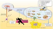
          Obesity is a public health issue that affects more than 600 million adults worldwide. The disease is characterized by fat accumulation, mainly in the abdominal area. The human body is mainly composed of two types of adipose tissue: white adipose tissue (WAT) and brown adipose tissue (BAT); however, the browning process generates a different type of brown fat-like adipocyte in WAT, which similar to BAT has thermogenic capacity by activating UCP-1. The hypothalamic arcuate nucleus plays an important role in WAT browning via POMC neurons, which are influenced by synergistic insulin and leptin signaling. On the other hand, stimulation of AgRP neurons suppresses WAT browning. The hypothalamic inflammatory process that occurs in obesity impairs insulin and leptin signaling in this tissue and, consequently, can decrease WAT browning. In addition, practicing physical exercise may be a great strategy for triggering the browning process since it reduces hypothalamic inflammation and increases POMC neurons gene expression. Moreover, physical exercise stimulates irisin gene expression, which has an important impact on thermogenesis, which in turn culminates in increased gene expression of proteins such as UCP-1 and Cidea, which are related to WAT browning. Furthermore, thermogenetic activation of WAT leads to increased energy expenditure, favoring obesity treatment. Therefore, this mini-review aimed to highlight the most recent studies that link the control of hypothalamic activity with the browning metabolism of adipose tissue in response to physical exercise.

          Related collections

          Most cited references31

          • Record: found
          • Abstract: found
          • Article: not found

          Exercise induces hippocampal BDNF through a PGC-1α/FNDC5 pathway.

          Exercise can improve cognitive function and has been linked to the increased expression of brain-derived neurotrophic factor (BDNF). However, the underlying molecular mechanisms driving the elevation of this neurotrophin remain unknown. Here we show that FNDC5, a previously identified muscle protein that is induced in exercise and is cleaved and secreted as irisin, is also elevated by endurance exercise in the hippocampus of mice. Neuronal Fndc5 gene expression is regulated by PGC-1α, and Pgc1a(-/-) mice show reduced Fndc5 expression in the brain. Forced expression of FNDC5 in primary cortical neurons increases Bdnf expression, whereas RNAi-mediated knockdown of FNDC5 reduces Bdnf. Importantly, peripheral delivery of FNDC5 to the liver via adenoviral vectors, resulting in elevated blood irisin, induces expression of Bdnf and other neuroprotective genes in the hippocampus. Taken together, our findings link endurance exercise and the important metabolic mediators, PGC-1α and FNDC5, with BDNF expression in the brain. Copyright © 2013 Elsevier Inc. All rights reserved.
            Bookmark
            • Record: found
            • Abstract: found
            • Article: not found

            Detection and Quantitation of Circulating Human Irisin by Tandem Mass Spectrometry.

            Exercise provides many health benefits, including improved metabolism, cardiovascular health, and cognition. We have shown previously that FNDC5, a type I transmembrane protein, and its circulating form, irisin, convey some of these benefits in mice. However, recent reports questioned the existence of circulating human irisin both because human FNDC5 has a non-canonical ATA translation start and because of claims that many human irisin antibodies used in commercial ELISA kits lack required specificity. In this paper we have identified and quantitated human irisin in plasma using mass spectrometry with control peptides enriched with heavy stable isotopes as internal standards. This precise state-of-the-art method shows that human irisin is mainly translated from its non-canonical start codon and circulates at ∼ 3.6 ng/ml in sedentary individuals; this level is increased to ∼ 4.3 ng/ml in individuals undergoing aerobic interval training. These data unequivocally demonstrate that human irisin exists, circulates, and is regulated by exercise.
              Bookmark
              • Record: found
              • Abstract: found
              • Article: not found

              Leptin and insulin act on POMC neurons to promote the browning of white fat.

              The primary task of white adipose tissue (WAT) is the storage of lipids. However, "beige" adipocytes also exist in WAT. Beige adipocytes burn fat and dissipate the energy as heat, but their abundance is diminished in obesity. Stimulating beige adipocyte development, or WAT browning, increases energy expenditure and holds potential for combating metabolic disease and obesity. Here, we report that insulin and leptin act together on hypothalamic neurons to promote WAT browning and weight loss. Deletion of the phosphatases PTP1B and TCPTP enhanced insulin and leptin signaling in proopiomelanocortin neurons and prevented diet-induced obesity by increasing WAT browning and energy expenditure. The coinfusion of insulin plus leptin into the CNS or the activation of proopiomelanocortin neurons also increased WAT browning and decreased adiposity. Our findings identify a homeostatic mechanism for coordinating the status of energy stores, as relayed by insulin and leptin, with the central control of WAT browning. Copyright © 2015 Elsevier Inc. All rights reserved.
                Bookmark

                Author and article information

                Contributors
                Journal
                Front Cell Neurosci
                Front Cell Neurosci
                Front. Cell. Neurosci.
                Frontiers in Cellular Neuroscience
                Frontiers Media S.A.
                1662-5102
                28 March 2018
                2018
                : 12
                : 88
                Affiliations
                [1] 1Laboratory of Molecular Biology of Exercise, School of Applied Sciences, University of Campinas , Limeira, Brazil
                [2] 2Department of Health Science, Federal University of Lavras , Lavras, Brazil
                [3] 3School of Physical Education and Sport of Ribeirão Preto, University of São Paulo , Ribeirão Preto, Brazil
                [4] 4Laboratory of Nutritional Genomics, School of Applied Sciences, University of Campinas , Limeira, Brazil
                [5] 5Center of Research in Sport Sciences, School of Applied Sciences, University of Campinas , Limeira, Brazil
                [6] 6University Catholic Center of Quixadá (Unicatolica) , Quixadá, Brazil
                [7] 7Postgraduate Program in Motricity Sciences, São Paulo State University , São Paulo, Brazil
                Author notes

                Edited by: Chao Deng, University of Wollongong, Australia

                Reviewed by: Miguel López, Universidade de Santiago de Compostela, Spain; Heike Muenzberg-Gruening, Pennington Biomedical Research Center, United States

                *Correspondence: Leandro P. de Moura leandropereiram@ 123456hotmail.com
                Article
                10.3389/fncel.2018.00088
                5882867
                8a93717e-8d83-4ecd-a5fa-f538f6263367
                Copyright © 2018 Rodrigues, Pereira, Campos, Moura, Silva, Cintra, Ropelle, Pauli, Araújo and Moura.

                This is an open-access article distributed under the terms of the Creative Commons Attribution License (CC BY). The use, distribution or reproduction in other forums is permitted, provided the original author(s) and the copyright owner are credited and that the original publication in this journal is cited, in accordance with accepted academic practice. No use, distribution or reproduction is permitted which does not comply with these terms.

                History
                : 03 August 2017
                : 13 March 2018
                Page count
                Figures: 1, Tables: 0, Equations: 0, References: 41, Pages: 7, Words: 5334
                Funding
                Funded by: Fundação de Amparo à Pesquisa do Estado de São Paulo 10.13039/501100001807
                Award ID: 2015/07199-2
                Award ID: 2016/24406-4
                Categories
                Neuroscience
                Mini Review

                Neurosciences
                wat browning,physical exercise,hypothalamus,pomc,agrp
                Neurosciences
                wat browning, physical exercise, hypothalamus, pomc, agrp

                Comments

                Comment on this article