4
views
0
recommends
+1 Recommend
0 collections
    0
    shares
      • Record: found
      • Abstract: found
      • Article: found
      Is Open Access

      Size-Dependent Cytotoxicity, Adhesion, and Endocytosis of Micro-/Nano-hydroxyapatite Crystals in HK-2 Cells

      research-article

      Read this article at

          There is no author summary for this article yet. Authors can add summaries to their articles on ScienceOpen to make them more accessible to a non-specialist audience.

          Abstract

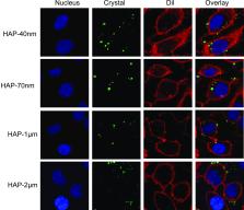
          Nano-hydroxyapatite (nano-HAP) is often used as a crystal nest to induce calcium oxalate (CaOx) kidney stone formation, but the mechanism of interaction between HAP crystals of different properties and renal tubular epithelial cells remains unclear. In this study, the adhesion and endocytosis of HAP crystals with sizes of 40 nm, 70 nm, 1 μm, and 2 μm (HAP-40 nm, HAP-70 nm, HAP-1 μm, and HAP-2 μm, respectively) to human renal proximal tubular epithelial cells (HK-2) were comparatively studied. The results showed that HAP crystals of all sizes promoted the expression of osteopontin and hyaluronic acid on the cell surface, destroyed the integrity of the lysosomes, and induced the apoptosis and necrosis of cells. Nano-HAP crystals had a higher specific surface area, a smaller contact angle, a higher surface energy, and a lower Zeta potential than those of micro-HAP. Therefore, the abilities of HK-2 cells to adhere to and endocytose nano-HAP crystals were greater than their abilities to do the same for micro-HAP crystals. The order of the endocytosed crystals was as follows: HAP-40 nm > HAP-70 nm > HAP-1 μm > HAP-2 μm. The endocytosed HAP crystals entered the lysosomes. The more crystal endocytosis and adhesion there is, the more toxic it is to HK-2 cells. The results of this study showed that nanosized HAP crystals greatly promoted the formation of kidney stones than micrometer-sized HAP crystals.

          Related collections

          Most cited references43

          • Record: found
          • Abstract: found
          • Article: not found

          Determining the size and shape dependence of gold nanoparticle uptake into mammalian cells.

          We investigated the intracellular uptake of different sized and shaped colloidal gold nanoparticles. We showed that kinetics and saturation concentrations are highly dependent upon the physical dimensions of the nanoparticles (e.g., uptake half-life of 14, 50, and 74 nm nanoparticles is 2.10, 1.90, and 2.24 h, respectively). The findings from this study will have implications in the chemical design of nanostructures for biomedical applications (e.g., tuning intracellular delivery rates and amounts by nanoscale dimensions and engineering complex, multifunctional nanostructures for imaging and therapeutics).
            Bookmark
            • Record: found
            • Abstract: found
            • Article: not found

            The effect of the shape of mesoporous silica nanoparticles on cellular uptake and cell function.

            The interaction between nanoparticles (NPs) and cells has been studied extensively, but the effect of particle shape on cell behavior has received little attention. Herein three different shaped monodisperse mesoporous silica nanoparticles (MSNs) of similar particle diameter, chemical composition and surface charge but with different aspect ratios (ARs, 1, 2, 4) were specially designed. Then the effects of particle shape of these three different shaped particles on cellular uptake and behavior were studied. The results indicated that these different shaped particles were readily internalized in A375 human melanoma (A375) cells by nonspecific cellular uptake. Particles with larger ARs were taken up in larger amounts and had faster internalization rates. Likewise, it was also found that particles with larger ARs had a greater impact on different aspects of cellular function including cell proliferation, apoptosis, cytoskeleton formation, adhesion and migration. These results show that nanoparticles should no longer be viewed as simple carriers for biomedical applications, but can also play an active role in mediating biological effects. Therefore, our findings may provide useful information for the development of new strategies for the design of efficient drug delivery nanocarriers and therapeutic systems and provide insights into nanotoxicity.
              Bookmark
              • Record: found
              • Abstract: not found
              • Article: not found

              Nanoparticle adhesion to the cell membrane and its effect on nanoparticle uptake efficiency.

              The interactions between nanosized particles and living systems are commonly mediated by what adsorbs to the nanoparticle in the biological environment, its biomolecular corona, rather than the pristine surface. Here, we characterize the adhesion toward the cell membrane of nanoparticles of different material and size and study how this is modulated by the presence or absence of a corona on the nanoparticle surface. The results are corroborated with adsorption to simple model supported lipid bilayers using a quartz crystal microbalance. We conclude that the adsorption of proteins on the nanoparticle surface strongly reduces nanoparticle adhesion in comparison to what is observed for the bare material. Nanoparticle uptake is described as a two-step process, where the nanoparticles initially adhere to the cell membrane and subsequently are internalized by the cells via energy-dependent pathways. The lowered adhesion in the presence of proteins thereby causes a concomitant decrease in nanoparticle uptake efficiency. The presence of a biomolecular corona may confer specific interactions between the nanoparticle-corona complex and the cell surface including triggering of regulated cell uptake. An important effect of the corona is, however, a reduction in the purely unspecific interactions between the bare material and the cell membrane, which in itself disregarding specific interactions, causes a decrease in cellular uptake. We suggest that future nanoparticle-cell studies include, together with characterization of size, charge, and dispersion stability, an evaluation of the adhesion properties of the material to relevant membranes.
                Bookmark

                Author and article information

                Journal
                ACS Omega
                ACS Omega
                ao
                acsodf
                ACS Omega
                American Chemical Society
                2470-1343
                05 December 2023
                19 December 2023
                : 8
                : 50
                : 48432-48443
                Affiliations
                []Department of Nephrology, The Second Affiliated Hospital, Xi’an Jiaotong University , Xi’an 710004, China
                []Department of Chemistry, Institute of Biomineralization and Lithiasis Research, Jinan University , Guangzhou, Guangdong 510632, China
                Author notes
                Author information
                https://orcid.org/0000-0001-8075-3915
                Article
                10.1021/acsomega.3c08180
                10733994
                38144057
                ae900172-4c08-40de-a15e-c247ef69af2a
                © 2023 The Authors. Published by American Chemical Society

                Permits non-commercial access and re-use, provided that author attribution and integrity are maintained; but does not permit creation of adaptations or other derivative works ( https://creativecommons.org/licenses/by-nc-nd/4.0/).

                History
                : 18 October 2023
                : 24 November 2023
                : 19 November 2023
                Funding
                Funded by: National Natural Science Foundation of China, doi 10.13039/501100001809;
                Award ID: 82270800
                Funded by: Zhongguancun Nephrology & Blood Purification Innovation Alliance, Youth Fund Project of CKD-MBD, doi NA;
                Award ID: NBPIA20QC0201
                Funded by: National Key Research and Development Program of China, doi 10.13039/501100012166;
                Award ID: 2020YFC2002700
                Categories
                Article
                Custom metadata
                ao3c08180
                ao3c08180

                Comments

                Comment on this article