3
views
0
recommends
+1 Recommend
0 collections
    0
    shares
      • Record: found
      • Abstract: found
      • Article: found
      Is Open Access

      ICG-Fluorescence Imaging for Margin Assessment During Minimally Invasive Colorectal Liver Metastasis Resection

      research-article
      , MD 1 , 2 , 3 , 4 , , MD 1 , 2 , 3 , 4 , , MD, PhD 2 , , MD, PhD 1 , , MD, PhD 3 , , MD, PhD 5 , , MD, PhD 6 , , MD, PhD 7 , , MD, PhD 8 , , MD, PhD 9 , , MD, PhD 10 , , MD, PhD 11 , , MD, PhD 12 , , MD, PhD 9 , , MD 3 , , MD 10 , 15 , , PhD 13 , , MD, PhD 1 , 14 , , MD, PhD 1 , , MD, PhD 1 , , MD, PhD 2 , 3 , 4 ,
      JAMA Network Open
      American Medical Association

      Read this article at

          There is no author summary for this article yet. Authors can add summaries to their articles on ScienceOpen to make them more accessible to a non-specialist audience.

          Key Points

          Question

          Is near-infrared fluorescence imaging with indocyanine green (ICG) associated with improved oncologic resections in patients undergoing minimally invasive resections of colorectal liver metastases?

          Findings

          In this cohort study of 201 adults with 316 histologically proven colorectal liver metastases, the overall rate of complete tumor resection was 92.4%. The absence of ICG fluorescence during parenchymal transection predicted a complete tumor resection (R0) with 92% accuracy.

          Meaning

          The findings of this study suggest that ICG fluorescence may provide surgeons with real-time feedback of the tumor margin during minimally invasive surgery for colorectal liver metastases and may increase the percentage of complete oncologic resections.

          Abstract

          Importance

          Unintended tumor-positive resection margins occur frequently during minimally invasive surgery for colorectal liver metastases and potentially negatively influence oncologic outcomes.

          Objective

          To assess whether indocyanine green (ICG)–fluorescence–guided surgery is associated with achieving a higher radical resection rate in minimally invasive colorectal liver metastasis surgery and to assess the accuracy of ICG fluorescence for predicting the resection margin status.

          Design, Setting, and Participants

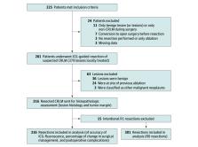
          The MIMIC (Minimally Invasive, Indocyanine-Guided Metastasectomy in Patients With Colorectal Liver Metastases) trial was designed as a prospective single-arm multicenter cohort study in 8 Dutch liver surgery centers. Patients were scheduled to undergo minimally invasive (laparoscopic or robot-assisted) resections of colorectal liver metastases between September 1, 2018, and June 30, 2021.

          Exposures

          All patients received a single intravenous bolus of 10 mg of ICG 24 hours prior to surgery. During surgery, ICG-fluorescence imaging was used as an adjunct to ultrasonography and regular laparoscopy to guide and assess the resection margin in real time. The ICG-fluorescence imaging was performed during and after liver parenchymal transection to enable real-time assessment of the tumor margin. Absence of ICG fluorescence was favorable both during transection and in the tumor bed directly after resection.

          Main Outcomes and Measures

          The primary outcome measure was the radical (R0) resection rate, defined by the percentage of colorectal liver metastases resected with at least a 1 mm distance between the tumor and resection plane. Secondary outcomes were the accuracy of ICG fluorescence in detecting margin-positive (R1; <1 mm margin) resections and the change in surgical management.

          Results

          In total, 225 patients were enrolled, of whom 201 (116 [57.7%] male; median age, 65 [IQR, 57-72] years) with 316 histologically proven colorectal liver metastases were included in the final analysis. The overall R0 resection rate was 92.4%. Re-resection of ICG-fluorescent tissue in the resection cavity was associated with a 5.0% increase in the R0 percentage (from 87.4% to 92.4%; P < .001). The sensitivity and specificity for real-time resection margin assessment were 60% and 90%, respectively (area under the receiver operating characteristic curve, 0.751; 95% CI, 0.668-0.833), with a positive predictive value of 54% and a negative predictive value of 92%. After training and proctoring of the first procedures, participating centers that were new to the technique had a comparable false-positive rate for predicting R1 resections during the first 10 procedures (odds ratio, 1.36; 95% CI, 0.44-4.24). The ICG-fluorescence imaging was associated with changes in intraoperative surgical management in 56 (27.9%) of the patients.

          Conclusions and Relevance

          In this multicenter prospective cohort study, ICG-fluorescence imaging was associated with an increased rate of tumor margin–negative resection and changes in surgical management in more than one-quarter of the patients. The absence of ICG fluorescence during liver parenchymal transection predicted an R0 resection with 92% accuracy. These results suggest that use of ICG fluorescence may provide real-time feedback of the tumor margin and a higher rate of complete oncologic resection.

          Abstract

          This cohort study of patients undergoing minimally invasive colorectal liver metastasis resections assesses the use of indocyanine green–fluorescence imaging to achieve a higher radical resection rate and greater accuracy in estimating the resection tumor-margin status.

          Related collections

          Most cited references40

          • Record: found
          • Abstract: not found
          • Article: not found

          World Medical Association Declaration of Helsinki: ethical principles for medical research involving human subjects.

          (2013)
            Bookmark
            • Record: found
            • Abstract: found
            • Article: not found

            Classification of surgical complications: a new proposal with evaluation in a cohort of 6336 patients and results of a survey.

            Although quality assessment is gaining increasing attention, there is still no consensus on how to define and grade postoperative complications. This shortcoming hampers comparison of outcome data among different centers and therapies and over time. A classification of complications published by one of the authors in 1992 was critically re-evaluated and modified to increase its accuracy and its acceptability in the surgical community. Modifications mainly focused on the manner of reporting life-threatening and permanently disabling complications. The new grading system still mostly relies on the therapy used to treat the complication. The classification was tested in a cohort of 6336 patients who underwent elective general surgery at our institution. The reproducibility and personal judgment of the classification were evaluated through an international survey with 2 questionnaires sent to 10 surgical centers worldwide. The new ranking system significantly correlated with complexity of surgery (P < 0.0001) as well as with the length of the hospital stay (P < 0.0001). A total of 144 surgeons from 10 different centers around the world and at different levels of training returned the survey. Ninety percent of the case presentations were correctly graded. The classification was considered to be simple (92% of the respondents), reproducible (91%), logical (92%), useful (90%), and comprehensive (89%). The answers of both questionnaires were not dependent on the origin of the reply and the level of training of the surgeons. The new complication classification appears reliable and may represent a compelling tool for quality assessment in surgery in all parts of the world.
              Bookmark
              • Record: found
              • Abstract: found
              • Article: not found

              Diagnosis and Treatment of Metastatic Colorectal Cancer : A Review

              Colorectal cancer (CRC) is the third most common cause of cancer mortality worldwide with more than 1.85 million cases and 850 000 deaths annually. Of new colorectal cancer diagnoses, 20% of patients have metastatic disease at presentation and another 25% who present with localized disease will later develop metastases.
                Bookmark

                Author and article information

                Journal
                JAMA Netw Open
                JAMA Netw Open
                JAMA Network Open
                American Medical Association
                2574-3805
                19 April 2024
                April 2024
                19 April 2024
                : 7
                : 4
                : e246548
                Affiliations
                [1 ]Department of Surgery, Leiden University Medical Center, Leiden, the Netherlands
                [2 ]Department of Surgery, Amsterdam University Medical Center, University of Amsterdam, Amsterdam, the Netherlands
                [3 ]Department of Surgery, Amsterdam University Medical Center, Vrije Universiteit Amsterdam, Amsterdam, the Netherlands
                [4 ]Cancer Center Amsterdam, Amsterdam, the Netherlands
                [5 ]NUTRIM School of Nutrition and Translational Research in Metabolism, Maastricht University Medical Center, Maastricht, the Netherlands
                [6 ]Department of Surgery, Jeroen Bosch Ziekenhuis, Den Bosch, the Netherlands
                [7 ]Department of Surgery, Maastricht University Medical Center, Maastricht, the Netherlands
                [8 ]Department of Surgery, St. Antonius Hospital, Nieuwegein/Regionaal Academisch Kankercentrum Utrecht, Utrecht, the Netherlands
                [9 ]Department of Surgery, Onze Lieve Vrouwe Gasthuis, Amsterdam, the Netherlands
                [10 ]Department of Surgery, Amphia Ziekenhuis, Breda, the Netherlands
                [11 ]Department of Surgery, University Medical Center Utrecht/Regionaal Academisch Kankercentrum Utrecht, Utrecht, the Netherlands
                [12 ]Department of Surgery, Medisch Spectrum Twente, Enschede, the Netherlands
                [13 ]Department of Medical Statistics and Bioinformatics, Leiden University Medical Center, Leiden, the Netherlands
                [14 ]Centre for Human Drug Research, Leiden, the Netherlands
                [15 ]Department of Surgical Oncology, Erasmus Medical Center, Rotterdam, the Netherlands
                Author notes
                Article Information
                Accepted for Publication: January 31, 2024.
                Published: April 19, 2024. doi:10.1001/jamanetworkopen.2024.6548
                Open Access: This is an open access article distributed under the terms of the CC-BY License. © 2024 Achterberg FB et al. JAMA Network Open.
                Corresponding Author: Rutger-Jan Swijnenburg, MD, PhD, Department of Surgery, Amsterdam University Medical Center, University of Amsterdam, De Boelelaan 1117, 1081 HV Amsterdam, the Netherlands ( r.j.swijnenburg@ 123456amsterdamumc.nl ).
                Author Contributions: Drs Achterberg and Bijlstra had full access to all of the data in the study and take responsibility for the integrity of the data and the accuracy of the data analysis. Drs Achterberg and Bijlstra served as co–first authors.
                Concept and design: Achterberg, Bijlstra, Sibinga Mulder, Bouwense, Coolsen, Mieog, Vahrmeijer, Swijnenburg.
                Acquisition, analysis, or interpretation of data: Achterberg, Bijlstra, Slooter, Boonstra, Bouwense, Bosscha, Derksen, Gerhards, Gobardhan, Hagendoorn, Lips, Marsman, Zonderhuis, Wullaert, Putter, Burggraaf, Mieog, Vahrmeijer, Swijnenburg.
                Drafting of the manuscript: Achterberg, Bijlstra, Bouwense, Swijnenburg.
                Critical review of the manuscript for important intellectual content: Achterberg, Bijlstra, Slooter, Sibinga Mulder, Boonstra, Bouwense, Bosscha, Coolsen, Derksen, Gerhards, Gobardhan, Hagendoorn, Lips, Marsman, Zonderhuis, Wullaert, Putter, Burggraaf, Mieog, Vahrmeijer, Swijnenburg.
                Statistical analysis: Achterberg, Bijlstra, Putter, Swijnenburg.
                Obtained funding: Swijnenburg.
                Administrative, technical, or material support: Achterberg, Bijlstra, Boonstra, Bouwense, Derksen, Hagendoorn, Lips, Marsman, Wullaert, Mieog, Vahrmeijer.
                Supervision: Bosscha, Derksen, Lips, Zonderhuis, Burggraaf, Mieog, Vahrmeijer, Swijnenburg.
                Conflict of Interest Disclosures: None reported.
                Funding/Support: This study was supported by grant 11289 from the KWF Kankerbestrijding (Dutch Cancer Society).
                Role of the Funder/Sponsor: The Dutch Cancer Society had no role in the design and conduct of the study; collection, management, analysis, and interpretation of the data; preparation, review, or approval of the manuscript; and decision to submit the manuscript for publication.
                Data Sharing Statement: See Supplement 2.
                Additional Contributions: We would like to acknowledge all patients, clinicians, and trial coordinators who participated in the trial.
                Article
                zoi240252
                10.1001/jamanetworkopen.2024.6548
                11031680
                38639939
                f49c8294-977a-4763-bca8-074e46a1175a
                Copyright 2024 Achterberg FB et al. JAMA Network Open.

                This is an open access article distributed under the terms of the CC-BY License.

                History
                : 9 October 2023
                : 31 January 2024
                Categories
                Research
                Original Investigation
                Online Only
                Surgery

                Comments

                Comment on this article