6
views
0
recommends
+1 Recommend
0 collections
    0
    shares
      • Record: found
      • Abstract: found
      • Article: found
      Is Open Access

      Rapid growth of a large penile median raphe cyst

      case-report

      Read this article at

          There is no author summary for this article yet. Authors can add summaries to their articles on ScienceOpen to make them more accessible to a non-specialist audience.

          Abstract

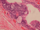
          A 57-year-old male presented to the emergency department due to sudden growth of a penile mass. On physical exam, the mass was located on the ventral surface of the penis at the level of the corona and measured 7cm × 4cm x 3.5cm. Ultrasound suggested that it was cystic in nature. The mass was surgically removed, and final pathology revealed a median raphe cyst.

          Related collections

          Most cited references4

          • Record: found
          • Abstract: found
          • Article: found
          Is Open Access

          Male median raphe cysts: serial retrospective analysis and histopathological classification

          Background To review the clinical and pathological characteristics of median raphe cysts and to classify the lesions according to pathogenesis and histopathological findings. Methods The medical records of patients who were diagnosed with median raphe cysts between 2001 and 2010 were reviewed to document the clinical presentation and pathological findings of the cysts. Results Most patients were asymptomatic; however, 9 patients had inflammatory or infectious cysts that were tender or painful. Four patients who had cysts on the parameatus and distal prepuce had difficulty voiding. Hematuria and hematospermia were noted in 2 cases. Thirty-one cysts were lined with an urothelium-like epithelium, and a squamous epithelium lining was found in 3 cases. In 2 cases, a well-formed mucinous glandular structure was observed. The other 20 cysts consisted of mixed epithelia. After excision of the cysts under local or general anesthesia, an urethral fistula developed as a complication in only 1 case. Conclusions Median raphe cysts are benign lesions formed due to tissue trapping during the development of urethral folds. The cysts can be defined into 4 types based on pathological findings: urethral, epidermoid, glandular, and mixed. The associated symptoms and signs should be taken into consideration when determining the treatment for the cysts. Virtual slides The virtual slide(s) for this article can be found here: http//http://www.diagnosticpathology.diagnomx.eu/vs/7727074877500751
            Bookmark
            • Record: found
            • Abstract: found
            • Article: found
            Is Open Access

            Median raphe cyst of the penis: a case report and review of the literature

            Background A defect in embryological development or closure of median raphe may lead to formation of cyst(s) anywhere in the midline from glans to anus. These cysts are referred to as median raphe cysts, an uncommonly encountered clinical condition. The cyst is generally solitary, with the penile shaft being the most common location, with average size of around 1 cm. The diagnosis is mostly clinical and confirmed histologically. We report a case of a patient with a rare histological variant of median raphe cyst and provide a focused review on presentation, histopathology, and management. Case presentation A 29-year-old unmarried Nepali man presented to our clinic with an asymptomatic, solitary, soft, translucent, nontender cystic lesion of about 1-cm diameter at the ventral aspect of glans penis, close to the meatus, that had been noticed at the age of 3 and was nonprogressive for the past 15 years. Ultrasonography demonstrated an isoechoic cystic lesion at the tip of the penis, separated from the urethra, and lying entirely within the mucosa without any evidence of solid component, septation, or vascularity. On the basis of clinical and ultrasonographic findings, a diagnosis of median raphe cyst of the penis was made. The cyst was excised with the patient under local anesthesia, and there was no evidence of recurrence in 2 years of follow-up. The histopathological examination with Hematoxylin and eosin staining showed the cyst wall was lined partly by ciliated pseudostratified columnar epithelium and partly by columnar epithelium with apical mucin. Conclusions Median raphe cyst is an uncommon, mostly asymptomatic condition in young patients. The cyst may occur anywhere along the midline from glans to anus. The diagnosis is clinical with histological confirmation. Excision is the treatment of choice with minimal chance of recurrence.
              Bookmark
              • Record: found
              • Abstract: found
              • Article: not found

              Penile Epidermal Cyst: A Case Report.

              Epidermal cysts also known as epidermoid cysts, is one of the common benign tumours presenting anywhere in the body. However, epidermal cyst in the penis is very rare. This condition in children is usually congenital due to abnormal embryologic closure of the median raphe; hence, it is termed as median raphe cysts (MRCs). Penile epidermal cysts may occur in adults following trauma or surgery due to epidermal elements being trapped within closed space. During wound healing, trapped squamous epithelium, undergoing keratinisation leads to cyst formation. Here, we report a rare case of patient with a penile epidermoid cyst whose main complaints was discomfort during coitus.
                Bookmark

                Author and article information

                Contributors
                Journal
                Urol Case Rep
                Urol Case Rep
                Urology Case Reports
                Elsevier
                2214-4420
                27 September 2021
                November 2021
                27 September 2021
                : 39
                : 101864
                Affiliations
                [a ]New York Medical College, 40 Sunshine Cottage Rd., Valhalla, NY, 10591, USA
                [b ]Pathology Assistant, Orange Pathology Associates, 707 East Main St., Middletown, NY, 10940, USA
                [c ]Chief of Anatomic Pathology, Good Samaritan Hospital, 255 Lafayette Ave., Suffern, NY, 10901, USA
                [d ]Bon Secours Medical Group, Division of Urology, Good Samaritan Hospital, 255 Lafayette Ave., Suffern, NY, 10901, USA
                Author notes
                []Corresponding author. smcnall@ 123456student.nymc.edu
                Article
                S2214-4420(21)00304-1 101864
                10.1016/j.eucr.2021.101864
                8488025
                04b07e16-9418-41ef-aa48-964b415b72b2
                © 2021 Published by Elsevier Inc.

                This is an open access article under the CC BY-NC-ND license (http://creativecommons.org/licenses/by-nc-nd/4.0/).

                History
                : 14 September 2021
                : 27 September 2021
                Categories
                Functional Urology

                median raphe cyst,penile cyst,penile mass,mrc, median raphe cyst

                Comments

                Comment on this article