4
views
0
recommends
+1 Recommend
0 collections
    0
    shares
      • Record: found
      • Abstract: found
      • Article: found
      Is Open Access

      Inflammatory cytokines as mediators of retinal endothelial barrier dysfunction in non‐infectious uveitis

      review-article

      Read this article at

          There is no author summary for this article yet. Authors can add summaries to their articles on ScienceOpen to make them more accessible to a non-specialist audience.

          Abstract

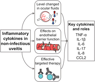
          Characterised by intraocular inflammation, non‐infectious uveitis includes a large group of autoimmune and autoinflammatory diseases that either involve the eye alone or have both ocular and systemic manifestations. When non‐infectious uveitis involves the posterior segment of the eye, specifically the retina, there is substantial risk of vision loss, often linked to breakdown of the inner blood‐retinal barrier. This barrier is formed by non‐fenestrated retinal vascular endothelial cells, reinforced by supporting cells that include pericytes, Müller cells and astrocytes. Across the published literature, a group of inflammatory cytokines stand out as prominent mediators of intraocular inflammation, with effects on the retinal endothelium that may contribute to breakdown of the inner blood‐retinal barrier, namely tumour necrosis factor (TNF)‐α, interleukin (IL)‐1β, IL‐6, IL‐8, IL‐17 and chemokine C‐C motif ligand (CCL)2. This article reviews the function of each cytokine and discusses the evidence for their involvement in retinal endothelial barrier dysfunction in non‐infectious uveitis, including basic laboratory investigations, studies of ocular fluids collected from patients with non‐infectious uveitis, and results of clinical treatment trials. The review also outlines gaps in knowledge in this area. Understanding the disease processes at a molecular level can suggest treatment alternatives that are directed against appropriate biological targets to protect the posterior segment of eye and preserve vision in non‐infectious uveitis.

          Abstract

          Non‐infectious uveitis has a range of ocular and systemic autoimmune and autoinflammatory causes. When the retina is involved, blood‐retinal barrier breakdown can lead to vision loss. Specific cytokines stand out as promoting intraocular inflammation. This article reviews the involvement of inflammatory cytokines – including tumour necrosis factor (TNF)‐α, interleukin (IL)‐1β, IL‐6, IL‐8, IL‐17 and chemokine C‐C motif ligand (CCL)2 – in non‐infectious uveitis. Their levels in ocular fluids, their effects on the endothelial barrier, and the results of clinical treatment trials are discussed.

          Related collections

          Most cited references259

          • Record: found
          • Abstract: found
          • Article: not found

          IL-6 in inflammation, immunity, and disease.

          Interleukin 6 (IL-6), promptly and transiently produced in response to infections and tissue injuries, contributes to host defense through the stimulation of acute phase responses, hematopoiesis, and immune reactions. Although its expression is strictly controlled by transcriptional and posttranscriptional mechanisms, dysregulated continual synthesis of IL-6 plays a pathological effect on chronic inflammation and autoimmunity. For this reason, tocilizumab, a humanized anti-IL-6 receptor antibody was developed. Various clinical trials have since shown the exceptional efficacy of tocilizumab, which resulted in its approval for the treatment of rheumatoid arthritis and juvenile idiopathic arthritis. Moreover, tocilizumab is expected to be effective for other intractable immune-mediated diseases. In this context, the mechanism for the continual synthesis of IL-6 needs to be elucidated to facilitate the development of more specific therapeutic approaches and analysis of the pathogenesis of specific diseases.
            Bookmark
            • Record: found
            • Abstract: found
            • Article: not found

            Standardization of Uveitis Nomenclature for Reporting Clinical Data. Results of the First International Workshop

            To begin a process of standardizing the methods for reporting clinical data in the field of uveitis. Consensus workshop. Members of an international working group were surveyed about diagnostic terminology, inflammation grading schema, and outcome measures, and the results used to develop a series of proposals to better standardize the use of these entities. Small groups employed nominal group techniques to achieve consensus on several of these issues. The group affirmed that an anatomic classification of uveitis should be used as a framework for subsequent work on diagnostic criteria for specific uveitic syndromes, and that the classification of uveitis entities should be on the basis of the location of the inflammation and not on the presence of structural complications. Issues regarding the use of the terms "intermediate uveitis," "pars planitis," "panuveitis," and descriptors of the onset and course of the uveitis were addressed. The following were adopted: standardized grading schema for anterior chamber cells, anterior chamber flare, and for vitreous haze; standardized methods of recording structural complications of uveitis; standardized definitions of outcomes, including "inactive" inflammation, "improvement'; and "worsening" of the inflammation, and "corticosteroid sparing," and standardized guidelines for reporting visual acuity outcomes. A process of standardizing the approach to reporting clinical data in uveitis research has begun, and several terms have been standardized.
              Bookmark
              • Record: found
              • Abstract: found
              • Article: not found

              The pro- and anti-inflammatory properties of the cytokine interleukin-6.

              Interleukin-6 is a cytokine not only involved in inflammation and infection responses but also in the regulation of metabolic, regenerative, and neural processes. In classic signaling, interleukin-6 stimulates target cells via a membrane bound interleukin-6 receptor, which upon ligand binding associates with the signaling receptor protein gp130. Gp130 dimerizes, leading to the activation of Janus kinases and subsequent phosphorylation of tyrosine residues within the cytoplasmic portion of gp130. This leads to the engagement of phosphatase Src homology domains containing tyrosin phosphatase-2 (SHP-2) and activation of the ras/raf/Mitogen-activated protein (MAP) kinase (MAPK) pathway. In addition, signal transducer and activator of transcription factors are recruited, which are phosphorylated, and consequently dimerize whereupon they translocate into the nucleus and activate target genes. Interestingly, only few cells express membrane bound interleukin-6 receptor whereas all cells display gp130 on the cell surface. While cells, which only express gp130, are not responsive to interleukin-6 alone, they can respond to a complex of interleukin-6 bound to a naturally occurring soluble form of the interleukin-6 receptor. Therefore, the generation of soluble form of the interleukin-6 receptor dramatically enlarges the spectrum of interleukin-6 target cells. This process has been named trans-signaling. Here, we review the involvement of both signaling modes in the biology of interleukin-6. It turns out that regenerative or anti-inflammatory activities of interleukin-6 are mediated by classic signaling whereas pro-inflammatory responses of interleukin-6 are rather mediated by trans-signaling. This is important since therapeutic blockade of interleukin-6 by the neutralizing anti-interleukin-6 receptor monoclonal antibody tocilizumab has recently been approved for the treatment of inflammatory diseases. This article is part of a Special Issue entitled: 11th European Symposium on Calcium. 2011 Elsevier B.V. All rights reserved.
                Bookmark

                Author and article information

                Contributors
                justine.smith@flinders.edu.au
                Journal
                Clin Transl Immunology
                Clin Transl Immunology
                10.1002/(ISSN)2050-0068
                CTI2
                Clinical & Translational Immunology
                John Wiley and Sons Inc. (Hoboken )
                2050-0068
                12 December 2023
                2023
                : 12
                : 12 ( doiID: 10.1002/cti2.v12.12 )
                : e1479
                Affiliations
                [ 1 ] Flinders University College of Medicine and Public Health Adelaide SA Australia
                Author notes
                [*] [* ] Correspondence

                JR Smith, Flinders University College of Medicine and Public Health, Flinders Medical Centre, Bedford Park, SA 5042, Australia.

                E‐mail: justine.smith@ 123456flinders.edu.au

                Author information
                https://orcid.org/0000-0002-4756-5493
                Article
                CTI21479 CTI-23-REV-0059.R2
                10.1002/cti2.1479
                10714664
                38090668
                4d65eab4-a040-42b7-ae1c-81ecb288cde7
                © 2023 The Authors. Clinical & Translational Immunology published by John Wiley & Sons Australia, Ltd on behalf of Australian and New Zealand Society for Immunology, Inc.

                This is an open access article under the terms of the http://creativecommons.org/licenses/by-nc-nd/4.0/ License, which permits use and distribution in any medium, provided the original work is properly cited, the use is non‐commercial and no modifications or adaptations are made.

                History
                : 22 September 2023
                : 17 November 2023
                : 19 April 2023
                : 21 November 2023
                Page count
                Figures: 1, Tables: 3, Pages: 22, Words: 19877
                Categories
                Review
                Review
                Custom metadata
                2.0
                2023
                Converter:WILEY_ML3GV2_TO_JATSPMC version:6.3.5 mode:remove_FC converted:12.12.2023

                cytokine,endothelial cell,endothelium,inflammation,retina,uveitis

                Comments

                Comment on this article