1
views
0
recommends
+1 Recommend
0 collections
    0
    shares
      • Record: found
      • Abstract: found
      • Article: found
      Is Open Access

      Reconstruction of the Esophagus with Sternohyoid Flap after Resection of a Large Cervical Esophageal Leiomyosarcoma

      case-report

      Read this article at

          There is no author summary for this article yet. Authors can add summaries to their articles on ScienceOpen to make them more accessible to a non-specialist audience.

          Abstract

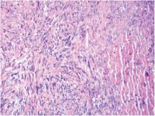
          Purpose: Reconstruction of the esophagus with sternohyoid muscle after enucleation of the cervical esophageal leiomyosarcoma (ELS) was rarely reported.

          Methods: A case of 55-year-old female with a large leiomyosarcoma in the cervical esophagus was reported. The tumor was enucleated, and the defect of the esophagus was patched with left sternohyoid muscle flap.

          Results: The patient recovered uneventfully after surgery. She has not had any discomfort with swallowing since surgery, and nowadays, there is not any recurrence and metastasis being detected.

          Conclusion: It is minimal invasive and simple to enucleate the cervical ELS and patch the defect of esophagus with sternohyoid muscle flap. For some selected patients, this method may be a promising surgical procedure to achieve both good swallowing function and satisfying prognosis.

          Related collections

          Most cited references10

          • Record: found
          • Abstract: found
          • Article: not found

          Esophageal sarcomas.

          The clinical and pathologic characteristics of five patients with esophageal sarcomas are presented, including the only recorded esophageal Triton tumor (malignant schwannoma), the third recorded synovial sarcoma, two patients with carcinosarcoma, and one with leiomyosarcoma. All five patients were males who presented with dysphagia. Three tumors were in the cervical esophagus, and the remaining two were in the distal esophagus. On endoscopic examination, three of the tumors were noted to be polypoid, while the other two were sessile. Two patients presented with disseminated disease, and a third was locally unresectable. These three patients were treated with palliative intent. The remaining two patients underwent surgical excision and postoperative radiation therapy, and are alive and well 6 and 7 years following treatment. This experience suggests that combined modality therapy employing postoperative radiation may be effective in managing these rare lesions.
            Bookmark
            • Record: found
            • Abstract: found
            • Article: not found

            Leiomyosarcoma of the esophagus: results of surgical treatment.

            This study examined the results of surgical treatment of leiomyosarcoma of the esophagus. Between January 1920 and December 1996, 17 patients (9 men and 8 women) with leiomyosarcoma of the esophagus were treated surgically at the Mayo Clinic. Median age was 58 years and ranged from 26 to 76 years. Symptoms included dysphagia in 11 patients (64.7%) and odynophagia in 6 (35.3%). The tumor was located in the middle third of the esophagus in 10 patients (58.8%) and in the cervical esophagus in 7 (41.2%). Procedures performed included esophagogastrectomy in 9 patients (Ivor Lewis in 5, left thoracoabdominal in 3, and transhiatal in 1), enucleation in 3, transgastric excision in 1, and exploration without resection in 4. The procedure was considered curative in 11 patients (64.7%). There was one operative death (mortality, 5.9%). Complications occurred in 3 patients (17.6%) and included anastomotic leak in 2 and bleeding requiring reoperation in 1. Growth pattern was infiltrating in 7, polypoid in 5, and intramural in 5. Histologically, the tumor was grade 1 in 6 patients, grade 2 in 2, grade 3 in 7, and grade 4 in 2. The tumor was postsurgically classified as stage I in 2 patients, stage IIA in 7, stage IIB in 1, stage IIIA in 5, stage IV in 1, and unknown in 1. Six patients (35.3%) received adjuvant treatment. Follow-up was complete in 16 patients (94.1%) and ranged from 1 to 182 months (median, 48 months). Five- and 10-year actuarial survivals were 47.0% and 31.0%, respectively. Seven patients (41.2%) are currently alive (median survival, 72 months); all underwent curative resection. Factors affecting survival included completeness of resection, growth pattern, postsurgical stage, tumor grade, and tumor location (p < 0.05). We conclude that leiomyosarcoma of the esophagus is rare. Complete resection provides long-term survival.
              Bookmark
              • Record: found
              • Abstract: found
              • Article: not found

              Esophageal leiomyosarcoma: clinical analysis and surgical treatment of 12 cases.

              Leiomyosarcoma of the esophagus is a rare malignant tumor with slow growth and late metastasis. The aim of this study was to reassess the clinical characteristics and treatment modality in one of the largest series of esophageal leiomyosarcomas from a single institution. From February 1973 to December 2011, 12 cases of esophageal leiomyosarcoma were identified. The principal symptoms included progressive dysphagia in 11 cases (91.7%), retrosternal/back pain in four (33.3%), weight loss in four (33.3%), upper gastrointestinal hemorrhage in two (16.7%), and emesis in two (16.7%). The average duration of symptoms was 10.6 months. The location of the primary tumor was in the middle thoracic esophagus in five cases, and lower thoracic esophagus in seven cases. Six cases were classified as the polypoid type, five cases as the infiltrative type, and only one case as the intramural type. All 12 of the patients underwent esophagectomies, and radical resections were achieved in these patients. Based on the Kaplan-Meier Method, the 3-, 5-, and 10-year survival rates were 80.0%, 58.3%, and 31.1%, respectively, with a median survival of 63 months. Five-year survival rates for patients with polypoid or intramural tumors (n = 7) was 83.3%, and for patients with infiltrative tumor (n = 5) it was 25.0%. One of the patients had tumor resected four times and survived for 161 months. In conclusion, patients presenting with esophageal leiomyosarcomas have an excellent prognosis, and radical resection may achieve acceptable results.
                Bookmark

                Author and article information

                Journal
                Ann Thorac Cardiovasc Surg
                Ann Thorac Cardiovasc Surg
                Annals of Thoracic and Cardiovascular Surgery
                The Editorial Committee of Annals of Thoracic and Cardiovascular Surgery
                1341-1098
                2186-1005
                13 December 2021
                2023
                : 29
                : 3
                : 148-152
                Affiliations
                [1 ]departmentDepartment of Thoracic Surgery , First Hospital of Tsinghua University , Beijing, China
                Author notes
                Corresponding author: Shenhai Wei, MD, PhD. Department of Thoracic Surgery, First Hospital of Tsinghua University, Beijing, 100016, China
                Article
                atcs.cr.21-00213
                10.5761/atcs.cr.21-00213
                10284660
                34897191
                97d905f5-abe7-437d-bcf3-d9090ccfcf31
                ©2023 Annals of Thoracic and Cardiovascular Surgery

                This work is licensed under a Creative Commons Attribution-NonCommercial-NoDerivatives International License

                History
                : 16 September 2021
                : 30 October 2021
                Categories
                Case Report

                esophageal leiomyosarcoma,surgical procedures,esophageal defect,sternohyoid muscle flap

                Comments

                Comment on this article