22
views
0
recommends
+1 Recommend
0 collections
    0
    shares
      • Record: found
      • Abstract: found
      • Article: found
      Is Open Access

      Myosin light-chain phosphatase regulates basal actomyosin oscillations during morphogenesis

      research-article

      Read this article at

          There is no author summary for this article yet. Authors can add summaries to their articles on ScienceOpen to make them more accessible to a non-specialist audience.

          Abstract

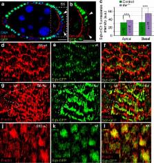
          Contractile actomyosin networks generate forces that drive tissue morphogenesis. Actomyosin contractility is controlled primarily by reversible phosphorylation of the myosin-II regulatory light chain through the action of myosin kinases and phosphatases. While the role of myosin light-chain kinase in regulating contractility during morphogenesis has been largely characterized, there is surprisingly little information on myosin light-chain phosphatase (MLCP) function in this context. Here, we use live imaging of Drosophila follicle cells combined with mathematical modelling to demonstrate that the MLCP subunit flapwing ( flw) is a key regulator of basal myosin oscillations and cell contractions underlying egg chamber elongation. Flw expression decreases specifically on the basal side of follicle cells at the onset of contraction and flw controls the initiation and periodicity of basal actomyosin oscillations. Contrary to previous reports, basal F-actin pulsates similarly to myosin. Finally, we propose a quantitative model in which periodic basal actomyosin oscillations arise in a cell-autonomous fashion from intrinsic properties of motor assemblies.

          Abstract

          Actomyosin contractility is regulated by phosphorylation of myosin regulatory light chain; much of the work in this area has focused on the kinase. Here the authors use Drosophila follicle cells and modelling to show that the phosphatase subunit Flapwing controls the initiation and dynamics of actomyosin oscillations.

          Related collections

          Most cited references43

          • Record: found
          • Abstract: found
          • Article: not found

          Non-muscle myosin II takes centre stage in cell adhesion and migration.

          Non-muscle myosin II (NM II) is an actin-binding protein that has actin cross-linking and contractile properties and is regulated by the phosphorylation of its light and heavy chains. The three mammalian NM II isoforms have both overlapping and unique properties. Owing to its position downstream of convergent signalling pathways, NM II is central in the control of cell adhesion, cell migration and tissue architecture. Recent insight into the role of NM II in these processes has been gained from loss-of-function and mutant approaches, methods that quantitatively measure actin and adhesion dynamics and the discovery of NM II mutations that cause monogenic diseases.
            Bookmark
            • Record: found
            • Abstract: found
            • Article: not found

            Planar polarized actomyosin contractile flows control epithelial junction remodelling.

            Force generation by Myosin-II motors on actin filaments drives cell and tissue morphogenesis. In epithelia, contractile forces are resisted at apical junctions by adhesive forces dependent on E-cadherin, which also transmits tension. During Drosophila embryonic germband extension, tissue elongation is driven by cell intercalation, which requires an irreversible and planar polarized remodelling of epithelial cell junctions. We investigate how cell deformations emerge from the interplay between force generation and cortical force transmission during this remodelling in Drosophila melanogaster. The shrinkage of dorsal-ventral-oriented ('vertical') junctions during this process is known to require planar polarized junctional contractility by Myosin II (refs 4, 5, 7, 12). Here we show that this shrinkage is not produced by junctional Myosin II itself, but by the polarized flow of medial actomyosin pulses towards 'vertical' junctions. This anisotropic flow is oriented by the planar polarized distribution of E-cadherin complexes, in that medial Myosin II flows towards 'vertical' junctions, which have relatively less E-cadherin than transverse junctions. Our evidence suggests that the medial flow pattern reflects equilibrium properties of force transmission and coupling to E-cadherin by α-Catenin. Thus, epithelial morphogenesis is not properly reflected by Myosin II steady state distribution but by polarized contractile actomyosin flows that emerge from interactions between E-cadherin and actomyosin networks.
              Bookmark
              • Record: found
              • Abstract: found
              • Article: not found

              Apical constriction: themes and variations on a cellular mechanism driving morphogenesis.

              Apical constriction is a cell shape change that promotes tissue remodeling in a variety of homeostatic and developmental contexts, including gastrulation in many organisms and neural tube formation in vertebrates. In recent years, progress has been made towards understanding how the distinct cell biological processes that together drive apical constriction are coordinated. These processes include the contraction of actin-myosin networks, which generates force, and the attachment of actin networks to cell-cell junctions, which allows forces to be transmitted between cells. Different cell types regulate contractility and adhesion in unique ways, resulting in apical constriction with varying dynamics and subcellular organizations, as well as a variety of resulting tissue shape changes. Understanding both the common themes and the variations in apical constriction mechanisms promises to provide insight into the mechanics that underlie tissue morphogenesis.
                Bookmark

                Author and article information

                Journal
                Nat Commun
                Nat Commun
                Nature Communications
                Nature Publishing Group
                2041-1723
                18 February 2016
                2016
                : 7
                : 10746
                Affiliations
                [1 ]Centro Andaluz de Biología del Desarrollo, Universidad Pablo de Olavide/CSIC/JA , Carretera de Utrera km 1, Sevilla 41013, Spain
                [2 ]Departamento de Física de la Materia Condensada, Instituto de Ciencias de Materiales Nicolás Cabrera, Condensed Matter Physics Center (IFIMAC), Universidad Autónoma de Madrid , Madrid 28049, Spain
                Author notes
                [*]

                These authors contributed equally to this work.

                Article
                ncomms10746
                10.1038/ncomms10746
                4759631
                26888436
                f6f38073-f39e-4a25-9b86-533173b4fcc7
                Copyright © 2016, Nature Publishing Group, a division of Macmillan Publishers Limited. All Rights Reserved.

                This work is licensed under a Creative Commons Attribution 4.0 International License. The images or other third party material in this article are included in the article's Creative Commons license, unless indicated otherwise in the credit line; if the material is not included under the Creative Commons license, users will need to obtain permission from the license holder to reproduce the material. To view a copy of this license, visit http://creativecommons.org/licenses/by/4.0/

                History
                : 13 November 2015
                : 18 January 2016
                Categories
                Article

                Uncategorized
                Uncategorized

                Comments

                Comment on this article