1
views
0
recommends
+1 Recommend
0 collections
    0
    shares
      • Record: found
      • Abstract: found
      • Article: found
      Is Open Access

      Distinct roles of ADIPOR1 and ADIPOR2: A pan-cancer analysis

      research-article

      Read this article at

          There is no author summary for this article yet. Authors can add summaries to their articles on ScienceOpen to make them more accessible to a non-specialist audience.

          Abstract

          Introduction

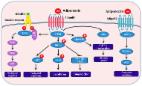
          AdipoR1 and AdipoR2 proteins, encoded by ADIPOR1 and ADIPOR2 genes respectively, are the receptors of adiponectin secrected by adipose tissue. Increasing studies have identified the vital role of adipose tissue in various diseases, including cancers. Hence, there is an urgent need to explore the roles of AdipoR1 and AdipoR2 in cancers.

          Methods

          We conducted a comprehensive pan-cancer analysis for the roles of AdipoR1 and AdipoR2 via several public databases, including expression differences, prognostic value, and the correlations with tumor microenvironment, epigenetic modification, and drug sensitivity.

          Results

          Both ADIPOR1 and ADIPOR2 genes are dysregulated in most cancers, but their genomic alteration frequencies are low. In addition, they are also correlated with the prognosis of some cancers. Although they are not strongly correlated with tumor mutation burden (TMB) or microsatellite instability (MSI), ADIPOR1/2 genes display a significant association with cancer stemness, tumor immune microenvironment, immune checkpoint genes (especially CD274 and NRP1), and drug sensitivity.

          Discussion

          ADIPOR1 and ADIPOR2 play critical roles in diverse cancers, and it is a potential strategy to treat tumors through targeting ADIPOR1 and ADIPOR2.

          Related collections

          Most cited references28

          • Record: found
          • Abstract: found
          • Article: not found

          Human Primary Liver Cancer -derived Organoid Cultures for disease modelling and drug screening

          Human liver cancer research currently lacks in vitro models that faithfully recapitulate the pathophysiology of the original tumour. We recently described a novel, near-physiological organoid culture system, where primary human healthy liver cells form long-term expanding organoids that retain liver tissue function and genetic stability. Here, we extend this culture system to the propagation of primary liver cancer (PLC) organoids from three of the most common PLC subtypes: hepatocellular carcinoma (HCC), cholangiocarcinoma (CC) and combined HCC/CC (CHC) tumours. PLC-derived organoid cultures preserve the histological architecture, gene expression and genomic landscape of the original tumour, allowing discrimination between different tumour tissues and subtypes, even after long term expansion in culture in the same medium conditions. Xenograft studies demonstrate that the tumourogenic potential, histological features and metastatic properties of PLC-derived organoids are preserved in vivo. PLC-derived organoids are amenable for biomarker identification and drug screening testing and lead to the identification of the ERK inhibitor SCH772984 as a potential therapeutic agent for primary liver cancer. We thus demonstrate the wide-ranging biomedical utilities of PLC-derived organoid models in furthering the understanding of liver cancer biology and in developing personalized medicine approaches for the disease.
            Bookmark
            • Record: found
            • Abstract: found
            • Article: found
            Is Open Access

            CancerSEA: a cancer single-cell state atlas

            Abstract High functional heterogeneity of cancer cells poses a major challenge for cancer research. Single-cell sequencing technology provides an unprecedented opportunity to decipher diverse functional states of cancer cells at single-cell resolution, and cancer scRNA-seq datasets have been largely accumulated. This emphasizes the urgent need to build a dedicated resource to decode the functional states of cancer single cells. Here, we developed CancerSEA (http://biocc.hrbmu.edu.cn/CancerSEA/ or http://202.97.205.69/CancerSEA/), the first dedicated database that aims to comprehensively explore distinct functional states of cancer cells at the single-cell level. CancerSEA portrays a cancer single-cell functional state atlas, involving 14 functional states (including stemness, invasion, metastasis, proliferation, EMT, angiogenesis, apoptosis, cell cycle, differentiation, DNA damage, DNA repair, hypoxia, inflammation and quiescence) of 41 900 cancer single cells from 25 cancer types. It allows querying which functional states are associated with the gene (or gene list) of interest in different cancers. CancerSEA also provides functional state-associated PCG/lncRNA repertoires across all cancers, in specific cancers, and in individual cancer single-cell datasets. In summary, CancerSEA provides a user-friendly interface for comprehensively searching, browsing, visualizing and downloading functional state activity profiles of tens of thousands of cancer single cells and the corresponding PCGs/lncRNAs expression profiles.
              Bookmark
              • Record: found
              • Abstract: found
              • Article: found
              Is Open Access

              Adiponectin, a Therapeutic Target for Obesity, Diabetes, and Endothelial Dysfunction

              Adiponectin is the most abundant peptide secreted by adipocytes, whose reduction plays a central role in obesity-related diseases, including insulin resistance/type 2 diabetes and cardiovascular disease. In addition to adipocytes, other cell types, such as skeletal and cardiac myocytes and endothelial cells, can also produce this adipocytokine. Adiponectin effects are mediated by adiponectin receptors, which occur as two isoforms (AdipoR1 and AdipoR2). Adiponectin has direct actions in liver, skeletal muscle, and the vasculature.Adiponectin exists in the circulation as varying molecular weight forms, produced by multimerization. Several endoplasmic reticulum ER-associated proteins, including ER oxidoreductase 1-α (Ero1-α), ER resident protein 44 (ERp44), disulfide-bond A oxidoreductase-like protein (DsbA-L), and glucose-regulated protein 94 (GPR94), have recently been found to be involved in the assembly and secretion of higher-order adiponectin complexes. Recent data indicate that the high-molecular weight (HMW) complexes have the predominant action in metabolic tissues. Studies have shown that adiponectin administration in humans and rodents has insulin-sensitizing, anti-atherogenic, and anti-inflammatory effects, and, in certain settings, also decreases body weight. Therefore, adiponectin replacement therapy in humans may suggest potential versatile therapeutic targets in the treatment of obesity, insulin resistance/type 2 diabetes, and atherosclerosis. The current knowledge on regulation and function of adiponectin in obesity, insulin resistance, and cardiovascular disease is summarized in this review.
                Bookmark

                Author and article information

                Contributors
                Journal
                Front Endocrinol (Lausanne)
                Front Endocrinol (Lausanne)
                Front. Endocrinol.
                Frontiers in Endocrinology
                Frontiers Media S.A.
                1664-2392
                21 February 2023
                2023
                : 14
                : 1119534
                Affiliations
                [1] 1 Central Laboratory of Yan’an Hospital Affiliated to Kunming Medical University , Kunming, Yunnan, China
                [2] 2 Department of Orthopedics of Yan’an Hospital Affiliated to Kunming Medical University , Kunming, Yunnan, China
                [3] 3 Greehey Children’s Cancer Research Institute, University of Texas Health at San Antonio , San Antonio, TX, United States
                Author notes

                Edited by: Bruno M. Simões, The University of Manchester, United Kingdom

                Reviewed by: Loredana Mauro, University of Calabria, Italy; Qianlei Zhou, Sun Yat-sen Memorial Hospital, China

                *Correspondence: Bin Yu, yubin100t@ 123456gmail.com

                This article was submitted to Cancer Endocrinology, a section of the journal Frontiers in Endocrinology

                Article
                10.3389/fendo.2023.1119534
                9990624
                36896172
                38f1cb3c-a0d8-4631-a2c8-b41a9af1c2d9
                Copyright © 2023 Chen, Yang, Ren, Yang, Huang, Li, Xiong and Yu

                This is an open-access article distributed under the terms of the Creative Commons Attribution License (CC BY). The use, distribution or reproduction in other forums is permitted, provided the original author(s) and the copyright owner(s) are credited and that the original publication in this journal is cited, in accordance with accepted academic practice. No use, distribution or reproduction is permitted which does not comply with these terms.

                History
                : 08 December 2022
                : 06 February 2023
                Page count
                Figures: 10, Tables: 0, Equations: 0, References: 28, Pages: 14, Words: 4149
                Categories
                Endocrinology
                Original Research

                Endocrinology & Diabetes
                adipose tissue,endocrine,adipor1,adipor2,tumor immune microenvironment,immunotherapy

                Comments

                Comment on this article