23
views
0
recommends
+1 Recommend
0 collections
    0
    shares
      • Record: found
      • Abstract: found
      • Article: found
      Is Open Access

      Comparing the Osteogenic Potentials and Bone Regeneration Capacities of Bone Marrow and Dental Pulp Mesenchymal Stem Cells in a Rabbit Calvarial Bone Defect Model

      research-article

      Read this article at

          There is no author summary for this article yet. Authors can add summaries to their articles on ScienceOpen to make them more accessible to a non-specialist audience.

          Abstract

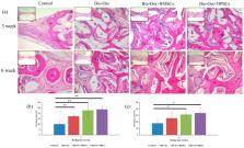
          The bone regeneration efficiency of bone marrow mesenchymal stem cells (BMSCs) and dental pulp mesenchymal stem cells (DPSCs) combined with xenografts in the craniofacial region remains unclear. Accordingly, this study commenced by comparing the cell morphology, cell proliferation, trilineage differentiation, mineral synthesis, and osteogenic gene expression of BMSCs and DPSCs in vitro. Four experimental groups (empty control, Bio-Oss only, Bio-Oss+BMSCs, and Bio-Oss+DPSCs) were then designed and implanted in rabbit calvarial defects. The BMSCs and DPSCs showed a similar morphology, proliferative ability, surface marker profile, and trilineage-differentiation potential in vitro. However, the BMSCs exhibited a higher mineral deposition and expression levels of osteogenic marker genes, including alkaline phosphatase (ALP), runt related transcription factor 2 (RUNX2), and osteocalcin (OCN). In the in vivo studies, the bone volume density in both MSC groups was significantly greater than that in the empty control or Bio-Oss only group. Moreover, the new bone formation and Collagen I / osteoprotegerin protein expressions of the scaffold+MSC groups were higher than those of the Bio-Oss only group. Finally, the Bio-Oss+BMSC and Bio-Oss+DPSC groups had a similar bone mineral density, new bone formation, and osteogenesis-related protein expression. Overall, the DPSCs seeded on Bio-Oss matched the bone regeneration efficacy of BMSCs in vivo and hence appear to be a promising strategy for craniofacial defect repair in future clinical applications.

          Related collections

          Most cited references38

          • Record: found
          • Abstract: found
          • Article: not found

          Mesenchymal stem cells: mechanisms of inflammation.

          In adults, human mesenchymal stem cells (hMSCs) are found in vivo at low frequency and are defined by their capacity to differentiate into bone, cartilage, and adipose tissue, depending on the stimuli and culture conditions under which they are expanded. Although MSCs were initially hypothesized to be the panacea for regenerating tissues, MSCs appear to be more important in therapeutics to regulate the immune response invoked in settings such as tissue injury, transplantation, and autoimmunity. MSCs have been used therapeutically in clinical trials and subsequently in practice to treat graft-versus-host disease following bone marrow transplantation. Reports of successful immune modulation suggest efficacy in a wide range of autoimmune conditions, such as demyelinating neurological disease (multiple sclerosis), systemic lupus erythematosus, and Crohn's disease, among others. This review provides background information about hMSCs and also describes their putative mechanisms of action in inflammation. We provide a summary of ongoing clinical trials to allow (a) full comprehension of the range of diseases in which hMSC therapy may be beneficial and (b) identification of gaps in our knowledge about the mechanisms of action of therapeutic MSCs in disease.
            Bookmark
            • Record: found
            • Abstract: found
            • Article: not found

            Concise reviews: Characteristics and potential applications of human dental tissue-derived mesenchymal stem cells.

            Recently, numerous types of human dental tissue-derived mesenchymal stem cells (MSCs) have been isolated and characterized, including dental pulp stem cells, stem cells from exfoliated deciduous teeth, periodontal ligament stem cells, dental follicle progenitor cells, alveolar bone-derived MSCs, stem cells from apical papilla, tooth germ progenitor cells, and gingival MSCs. All these MSC-like cells exhibit self-renewal, multilineage differentiation potential, and immunomodulatory properties. Several studies have demonstrated the potential advantages of dental stem cell-based approaches for regenerative treatments and immunotherapies. This review outlines the properties of various dental MSC-like populations and the progress toward their use in regenerative therapy. Several dental stem cell banks worldwide are also introduced, with a view toward future clinical application.
              Bookmark
              • Record: found
              • Abstract: found
              • Article: found
              Is Open Access

              Tissue source determines the differentiation potentials of mesenchymal stem cells: a comparative study of human mesenchymal stem cells from bone marrow and adipose tissue

              Background Mesenchymal stem cells (MSCs) possess intrinsic regeneration capacity as part of the repair process in response to injury, such as fracture or other tissue injury. Bone marrow and adipose tissue are the major sources of MSCs. However, which cell type is more effective and suitable for cell therapy remains to be answered. The intrinsic molecular mechanism supporting the assertion has also been lacking. Methods Human bone marrow-derived MSCs (BMSCs) and adipose tissue-derived MSCs (ATSCs) were isolated from bone marrow and adipose tissue obtained after total hip arthroplasty. ATSCs and BMSCs were incubated in standard growth medium. Trilineage differentiation including osteogenesis, adipogenesis, and chondrogenesis was performed by addition of relevant induction mediums. The expression levels of trilineage differentiation marker genes were evaluated by quantitative RT-PCR. The methylation status of CpG sites of Runx2, PPARγ, and Sox9 promoters were checked by bisulfite sequencing. In addition, ectopic bone formation and calvarial bone critical defect models were used to evaluate the bone regeneration ability of ATSCs and BMSCs in vivo. Results The results showed that BMSCs possessed stronger osteogenic and lower adipogenic differentiation potentials compared to ATSCs. There was no significant difference in the chondrogenic differentiation potential. The CpG sites of Runx2 promoter in BMSCs were hypomethylated, while in ATSCs they were hypermethylated. The CpG sites of PPARγ promoter in ATSCs were hypomethylated, while in BMSCs they were hypermethylated. The methylation status of Sox9 promoter in BMSCs was only slightly lower than that in ATSCs. Conclusions The epigenetic memory obtained from either bone marrow or adipose tissue favored MSC differentiation along an osteoblastic or adipocytic lineage. The methylation status of the main transcription factors controlling MSC fate contributes to the differential differentiation capacities of different source-derived MSCs. Electronic supplementary material The online version of this article (doi:10.1186/s13287-017-0716-x) contains supplementary material, which is available to authorized users.
                Bookmark

                Author and article information

                Journal
                Int J Mol Sci
                Int J Mol Sci
                ijms
                International Journal of Molecular Sciences
                MDPI
                1422-0067
                10 October 2019
                October 2019
                : 20
                : 20
                : 5015
                Affiliations
                [1 ]Department of Obstetrics and Gynecology, Taipei Medical University Hospital, Taipei 110, Taiwan; d119097012@ 123456tmu.edu.tw
                [2 ]School of Dentistry, College of Oral Medicine, Taipei Medical University, Taipei 110, Taiwan; babypg666666@ 123456gmail.com (Y.-H.C.); endo@ 123456tmu.edu.tw (S.-C.H.); b202094090@ 123456tmu.edu.tw (W.-Z.L.)
                [3 ]School of Oral Hygiene, College of Oral Medicine, Taipei Medical University, Taipei 110, Taiwan
                [4 ]Division of Prosthodontics, Department of Dentistry, Taipei Medical University Hospital, Taipei 110, Taiwan
                Author notes
                [* ]Correspondence: shengwei@ 123456tmu.edu.tw ; Tel.: +886-2-2736-1661 (ext. 5401); Fax: +886-2-27362295
                [†]

                Authors contributed equally to this manuscript.

                Author information
                https://orcid.org/0000-0001-6637-7044
                Article
                ijms-20-05015
                10.3390/ijms20205015
                6834129
                31658685
                5ffa9da9-e21e-4269-b98d-d79a11e42607
                © 2019 by the authors.

                Licensee MDPI, Basel, Switzerland. This article is an open access article distributed under the terms and conditions of the Creative Commons Attribution (CC BY) license ( http://creativecommons.org/licenses/by/4.0/).

                History
                : 05 September 2019
                : 09 October 2019
                Categories
                Article

                Molecular biology
                mesenchymal stem cells,dental pulp stem cells,bone marrow stem cells,bone regeneration,calvarial defect

                Comments

                Comment on this article