1
views
0
recommends
+1 Recommend
0 collections
    0
    shares
      • Record: found
      • Abstract: found
      • Article: found
      Is Open Access

      Changes in Glial Fibrillary Acidic Protein-Immunoreactive Astrocytes in the Prefrontal Cortex of the Male Rat following Chronic Khat Use

      research-article

      Read this article at

          There is no author summary for this article yet. Authors can add summaries to their articles on ScienceOpen to make them more accessible to a non-specialist audience.

          Abstract

          Background:

          Long-term khat consumption is associated with significant neurocognitive changes, which have been elucidated in behavioral studies. With current research showing the centrality of astrocytes and other glial cells in neuronal signaling, there is possibility that these cells are also affected by chronic khat use. There is little literature on the structural changes in the prefrontal cortex neuronal and astrocytic cytoarchitecture and morphometry in chronic khat users.

          Objective:

          The objective of this study was to describe the changes in astrocyte morphometry and structure in rats after long-term use of khat (miraa).

          Materials and Methods:

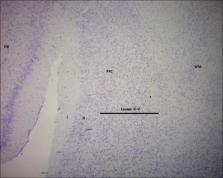
          Adult male Wistar rats, aged 2–3 months, weighing 200–300 g were randomized into four groups of 10 each (control, Group 1, Group 2, and Group 3) to correspond with those used as controls and those that received 500 mg/kg, 1000 mg/kg, and 2000 mg/kg body weight khat extracts, respectively. Fresh khat leaves were purchased from Maua market in Meru, and crude extract was prepared using lyophilization. The control rats were fed on normal diet, while the experimental groups were fed on normal diet and khat extracts using oral gavage for 6 weeks. The animals were sacrificed and their brains were removed. We performed immunohistochemical visualization of astrocytes using glial fibrillary acidic protein. Photomicrographs of the stained sections were transferred to ImageJ Fiji software to study the astrocyte density and astrocytic processes. We used Kruskal–Wallis test to correlate the four animal groups in terms of astrocyte densities.

          Results:

          We observed an increase in the average number of astrocytes with increasing doses of khat compared to controls, with those in Group 3 (2000 mg/kg) having an exuberant reactive astrocytosis. Further, escalating khat doses resulted in increased glial fibrillary acidic protein immunoreactivity in the nuclei and astrocytic processes, gliotic changes, and increased complexity of astrocytic processes.

          Conclusion:

          Chronic khat use, especially at high doses, results in reactive astrocytosis and astrogliosis, which may be part of the mechanisms involved in the cognitive changes associated with its use.

          Related collections

          Most cited references38

          • Record: found
          • Abstract: found
          • Article: not found

          Astrocytes: biology and pathology

          Astrocytes are specialized glial cells that outnumber neurons by over fivefold. They contiguously tile the entire central nervous system (CNS) and exert many essential complex functions in the healthy CNS. Astrocytes respond to all forms of CNS insults through a process referred to as reactive astrogliosis, which has become a pathological hallmark of CNS structural lesions. Substantial progress has been made recently in determining functions and mechanisms of reactive astrogliosis and in identifying roles of astrocytes in CNS disorders and pathologies. A vast molecular arsenal at the disposal of reactive astrocytes is being defined. Transgenic mouse models are dissecting specific aspects of reactive astrocytosis and glial scar formation in vivo. Astrocyte involvement in specific clinicopathological entities is being defined. It is now clear that reactive astrogliosis is not a simple all-or-none phenomenon but is a finely gradated continuum of changes that occur in context-dependent manners regulated by specific signaling events. These changes range from reversible alterations in gene expression and cell hypertrophy with preservation of cellular domains and tissue structure, to long-lasting scar formation with rearrangement of tissue structure. Increasing evidence points towards the potential of reactive astrogliosis to play either primary or contributing roles in CNS disorders via loss of normal astrocyte functions or gain of abnormal effects. This article reviews (1) astrocyte functions in healthy CNS, (2) mechanisms and functions of reactive astrogliosis and glial scar formation, and (3) ways in which reactive astrocytes may cause or contribute to specific CNS disorders and lesions.
            Bookmark
            • Record: found
            • Abstract: found
            • Article: not found

            Leukocyte infiltration, neuronal degeneration, and neurite outgrowth after ablation of scar-forming, reactive astrocytes in adult transgenic mice.

            Reactive astrocytes adjacent to a forebrain stab injury were selectively ablated in adult mice expressing HSV-TK from the Gfap promoter by treatment with ganciclovir. Injured tissue that was depleted of GFAP-positive astrocytes exhibited (1) a prolonged 25-fold increase in infiltration of CD45-positive leukocytes, including ultrastructurally identified monocytes, macrophages, neutrophils, and lymphocytes, (2) failure of blood-brain barrier (BBB) repair, (3) substantial neuronal degeneration that could be attenuated by chronic glutamate receptor blockade, and (4) a pronounced increase in local neurite outgrowth. These findings show that genetic targeting can be used to ablate scar-forming astrocytes and demonstrate roles for astrocytes in regulating leukocyte trafficking, repairing the BBB, protecting neurons, and restricting nerve fiber growth after injury in the adult central nervous system.
              Bookmark
              • Record: found
              • Abstract: found
              • Article: not found

              Coordinate regulation of glutathione biosynthesis and release by Nrf2-expressing glia potently protects neurons from oxidative stress.

              Astrocytes have a higher antioxidant potential in comparison to neurons. Pathways associated with this selective advantage include the transcriptional regulation of antioxidant enzymes via the action of the Cap'n'Collar transcription factor Nrf2 at the antioxidant response element (ARE). Here we show that Nrf2 overexpression can reengineer neurons to express this glial pathway and enhance antioxidant gene expression. However, Nrf2-mediated protection from oxidative stress is conferred primarily by glia in mixed cultures. The antioxidant properties of Nrf2-overexpressing glia are more pronounced than those of neurons, and a relatively small number of these glia (< 1% of total cell number added) could protect fully cocultured naive neurons from oxidative glutamate toxicity associated with glutathione (GSH) depletion. Microarray and biochemical analyses indicate a coordinated upregulation of enzymes involved in GSH biosynthesis (xCT cystine antiporter, gamma-glutamylcysteine synthetase, and GSH synthase), use (glutathione S-transferase and glutathione reductase), and export (multidrug resistance protein 1) with Nrf2 overexpression, leading to an increase in both media and intracellular GSH. Selective inhibition of glial GSH synthesis and the supplementation of media GSH indicated that an Nrf2-dependent increase in glial GSH synthesis was both necessary and sufficient for the protection of neurons, respectively. Neuroprotection was not limited to overexpression of Nrf2, because activation of endogenous glial Nrf2 by the small molecule ARE inducer, tert-butylhydroquinone, also protected against oxidative glutamate toxicity.
                Bookmark

                Author and article information

                Journal
                J Microsc Ultrastruct
                J Microsc Ultrastruct
                JMAU
                J Microsc Ultrastruct
                Journal of Microscopy and Ultrastructure
                Wolters Kluwer - Medknow (India )
                2213-879X
                2213-8803
                Apr-Jun 2024
                07 February 2023
                : 12
                : 2
                : 75-80
                Affiliations
                [1 ]Department of Human Anatomy, School of Medicine and Health Sciences, Kenya Methodist University, Meru, Nairobi, Kenya
                [2 ]Department of Human Anatomy and Physiology, Faculty of Health Sciences, The University of Nairobi, Nairobi, Kenya
                [3 ]Department of Morphological Sciences, School of Anatomical Sciences, Faculty of Health Sciences, University of the Witwatersrand Johannesburg, South Africa
                [4 ]Department of Anatomy, Johannesburg University, Johannesburg, South Africa
                [5 ]Department of Quality Assurance, Kenya Bureau of Standards, Nairobi, Kenya
                Author notes
                Address for correspondence: Dr. Paul Bundi Karau, Department of Human Anatomy, School of Medicine, Kenya Methodist University, P. O. Box 2375-60200, Meru, Kenya. E-mail: pbkarau@ 123456gmail.com
                Article
                JMAU-12-75
                10.4103/jmau.jmau_26_22
                11245132
                39006043
                a09dbf46-bb93-4ada-b24f-17e01835cbd7
                Copyright: © 2023 Journal of Microscopy and Ultrastructure

                This is an open access journal, and articles are distributed under the terms of the Creative Commons Attribution-NonCommercial-ShareAlike 4.0 License, which allows others to remix, tweak, and build upon the work non-commercially, as long as appropriate credit is given and the new creations are licensed under the identical terms.

                History
                : 23 March 2022
                : 17 May 2022
                : 23 May 2022
                Categories
                Original Article

                chronic consumption,glial fibrillary acidic protein,khat,reactive astrocytes

                Comments

                Comment on this article