178
views
0
recommends
+1 Recommend
0 collections
    0
    shares
      • Record: found
      • Abstract: found
      • Article: found
      Is Open Access

      A bioprosthetic ovary created using 3D printed microporous scaffolds restores ovarian function in sterilized mice

      research-article

      Read this article at

          There is no author summary for this article yet. Authors can add summaries to their articles on ScienceOpen to make them more accessible to a non-specialist audience.

          Abstract

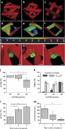
          Emerging additive manufacturing techniques enable investigation of the effects of pore geometry on cell behavior and function. Here, we 3D print microporous hydrogel scaffolds to test how varying pore geometry, accomplished by manipulating the advancing angle between printed layers, affects the survival of ovarian follicles. 30° and 60° scaffolds provide corners that surround follicles on multiple sides while 90° scaffolds have an open porosity that limits follicle–scaffold interaction. As the amount of scaffold interaction increases, follicle spreading is limited and survival increases. Follicle-seeded scaffolds become highly vascularized and ovarian function is fully restored when implanted in surgically sterilized mice. Moreover, pups are born through natural mating and thrive through maternal lactation. These findings present an in vivo functional ovarian implant designed with 3D printing, and indicate that scaffold pore architecture is a critical variable in additively manufactured scaffold design for functional tissue engineering.

          Abstract

          There is a clinical need to develop a bioengineering system to support ovary transplantation. Here, the authors generate a bioprosthetic ovary using 3D printed scaffolds of varying pore architectures to support follicle survival and ovarian function in sterilized mice.

          Related collections

          Most cited references59

          • Record: found
          • Abstract: found
          • Article: not found

          Three-dimensional bioprinting of thick vascularized tissues.

          The advancement of tissue and, ultimately, organ engineering requires the ability to pattern human tissues composed of cells, extracellular matrix, and vasculature with controlled microenvironments that can be sustained over prolonged time periods. To date, bioprinting methods have yielded thin tissues that only survive for short durations. To improve their physiological relevance, we report a method for bioprinting 3D cell-laden, vascularized tissues that exceed 1 cm in thickness and can be perfused on chip for long time periods (>6 wk). Specifically, we integrate parenchyma, stroma, and endothelium into a single thick tissue by coprinting multiple inks composed of human mesenchymal stem cells (hMSCs) and human neonatal dermal fibroblasts (hNDFs) within a customized extracellular matrix alongside embedded vasculature, which is subsequently lined with human umbilical vein endothelial cells (HUVECs). These thick vascularized tissues are actively perfused with growth factors to differentiate hMSCs toward an osteogenic lineage in situ. This longitudinal study of emergent biological phenomena in complex microenvironments represents a foundational step in human tissue generation.
            Bookmark
            • Record: found
            • Abstract: found
            • Article: not found

            Direct 3D Printing of Shear-Thinning Hydrogels into Self-Healing Hydrogels.

            Supramolecular hydrogels are used in the 3D printing of high-resolution, multi-material structures. The non-covalent bonds allow the extrusion of the inks into support gels to directly write structures continuously in 3D space. This material system supports the patterning of multiple inks, cells, and void spaces.
              Bookmark
              • Record: found
              • Abstract: found
              • Article: not found

              Gelatin as a delivery vehicle for the controlled release of bioactive molecules.

              Gelatin is a commonly used natural polymer which is derived from collagen. The isoelectric point of gelatin can be modified during the fabrication process to yield either a negatively charged acidic gelatin, or a positively charged basic gelatin at physiological pH. This theoretically allows electrostatic interactions to take place between a charged biomolecule and gelatin of the opposite charge, forming polyion complexes. Various forms of gelatin carrier matrices can be fabricated for controlled-release studies, and characterization studies have been performed which show that gelatin carriers are able to sorb charged biomolecules such as proteins and plasmid DNA through polyion complexation. The crosslinking density of gelatin hydrogels has been shown to affect their degradation rate in vivo, and the rate of biomolecule release from gelatin carriers has been shown to have a similar profile, suggesting that complexed gelatin/biomolecule fragments are released by enzymatic degradation of the carrier in vivo. This review will emphasize how biomolecules released from gelatin controlled-release systems are able to retain their biological activity, allowing for their use in tissue engineering, therapeutic angiogenesis, gene therapy, and drug delivery applications.
                Bookmark

                Author and article information

                Journal
                Nat Commun
                Nat Commun
                Nature Communications
                Nature Publishing Group
                2041-1723
                16 May 2017
                2017
                : 8
                : 15261
                Affiliations
                [1 ]Division of Reproductive Biology in Medicine, Department of Obstetrics and Gynecology, Feinberg School of Medicine, Northwestern University , Chicago, Illinois 60611, USA
                [2 ]Center for Reproductive Science, Northwestern University , Chicago, Illinois 60611, USA
                [3 ]Oncofertility Consortium, Northwestern University , Chicago, Illinois 60611, USA
                [4 ]Simpson Querrey Institute for BioNanotechnology, Northwestern University , Chicago, Illinois 60611, USA
                [5 ]Department of Biomedical Engineering, Northwestern University , Evanston, Illinois 60208, USA
                [6 ]Department of Anatomy and Cell Biology, University of Kansas Medical Center , Kansas City, Kansas 66160, USA
                [7 ]Northwestern University Atomic and Nanoscale Characterization Experimental Center, Northwestern University , Evanston, Illinois 60208, USA
                [8 ]Department of Materials Science and Engineering, Northwestern University , Evanston, Illinois 60208, USA
                [9 ]Department of Surgery, Feinberg School of Medicine, Northwestern University , Chicago, Illinois 60611, USA
                Author notes
                [*]

                These authors contributed equally to this work.

                Author information
                http://orcid.org/0000-0001-7840-4695
                http://orcid.org/0000-0002-1197-3399
                Article
                ncomms15261
                10.1038/ncomms15261
                5440811
                28509899
                1389d1cb-8e78-4d54-a50c-0c98cba17c2b
                Copyright © 2017, The Author(s)

                This work is licensed under a Creative Commons Attribution 4.0 International License. The images or other third party material in this article are included in the article's Creative Commons license, unless indicated otherwise in the credit line; if the material is not included under the Creative Commons license, users will need to obtain permission from the license holder to reproduce the material. To view a copy of this license, visit http://creativecommons.org/licenses/by/4.0/

                History
                : 09 February 2017
                : 14 March 2017
                Categories
                Article

                Uncategorized
                Uncategorized

                Comments

                Comment on this article