3
views
0
recommends
+1 Recommend
0 collections
    0
    shares
      • Record: found
      • Abstract: found
      • Article: found
      Is Open Access

      Reference ranges for the fetal mesencephalon to occiput measurement at 11 to 13+6 weeks of gestation

      research-article

      Read this article at

          There is no author summary for this article yet. Authors can add summaries to their articles on ScienceOpen to make them more accessible to a non-specialist audience.

          Abstract

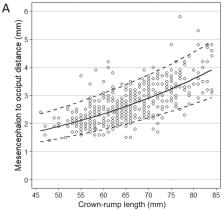
          The objective was to have a quantitative description of the normal position of the fetal midbrain in the first trimester, through defining the reference ranges for the mesencephalon to the occipital bone distance, in the axial plane. This was a prospective study that included normal fetuses screened between 11 and 13 weeks of gestation. The distance was measured between the posterior limit of the mesencephalon to the occipital bone in the same axial view as the one required for the biparietal diameter (BPD) assessment, at this gestational age (GA). The reference ranges using quantile regression, according to the crown-rump length (CRL), BPD, and GA were fitted. Data analysis included 428 ultrasound measurements. A good, linear correlation was observed between mesencephalon to occiput (MO) distance and CRL, BPD, or GA. It increased linearly with advancing gestation (log 10MO = -0.1834 + 0.0092 x CRL, R 2=0.48, P<0.0001) and was independent of maternal demographic characteristics and intracranial translucency (IT). In our study, the 1st percentile of the normal MO distance varies from 1.31 mm at a CRL of 45 mm to 2.08 mm at a CRL of 84 mm. The intraclass correlation coefficient (ICC) was 0.89 for intraobserver variability. A significant increase in the MO distance was found in the patients who did not receive folic acid in the first trimester of pregnancy [1.056 vs. 1.008 multiple of median (MoM), P=0.014]. A simple measurement is described between the midbrain and the occipital bone, obtained in the same axial view. It increases linearly with advancing gestation. Integration of this measurement into the routine ultrasound screening in association with the ‘crash sign’ and recognizing the lower extreme values could lead to an early diagnosis of open spina bifida (OSB).

          Related collections

          Most cited references19

          • Record: found
          • Abstract: not found
          • Article: not found

          ISUOG practice guidelines: performance of first-trimester fetal ultrasound scan.

            Bookmark
            • Record: found
            • Abstract: found
            • Article: not found

            New charts for ultrasound dating of pregnancy and assessment of fetal growth: longitudinal data from a population-based cohort study.

            Correct assessment of gestational age and fetal growth is essential for optimal obstetric management. The objectives of this study were, first, to develop charts for ultrasound dating of pregnancy based on crown-rump length and biparietal diameter and, second, to derive reference curves for normal fetal growth based on biparietal diameter, head circumference, transverse cerebellar diameter, abdominal circumference and femur length from 10 weeks of gestational age onwards. A total of 8313 pregnant women were included for analysis in this population-based prospective cohort study. All women had repeated ultrasound assessments to examine fetal growth. Charts for ultrasound dating of pregnancy, based on crown-rump length and biparietal diameter, were derived. Internal validation with the actual date of delivery showed that ultrasound imaging provided reliable gestational age estimates. Up to 92% of deliveries took place within 37-42 weeks of gestation if gestational age was derived from ultrasound data, compared with 87% based on a reliable last menstrual period. The earlier the ultrasound assessment the more accurate the prediction of date of delivery. After 24 weeks of gestation a reliable last menstrual period provided better estimates of gestational age. Reference curves for normal fetal growth from 10 weeks of gestational age onwards were derived. Charts for ultrasound dating of pregnancy and reference curves for fetal biometry are presented. The results indicate that, up to 24 weeks of pregnancy, dating by ultrasound examination provides a better prediction of the date of delivery than does last menstrual period. The earlier the ultrasound assessment in pregnancy, preferably between 10 and 12 weeks, the better the estimate of gestational age. Copyright (c) 2008 ISUOG. Published by John Wiley & Sons, Ltd.
              Bookmark
              • Record: found
              • Abstract: found
              • Article: not found

              Assessment of intracranial translucency (IT) in the detection of spina bifida at the 11-13-week scan.

              Prenatal diagnosis of open spina bifida is carried out by ultrasound examination in the second trimester of pregnancy. The diagnosis is suspected by the presence of a 'lemon-shaped' head and a 'banana-shaped' cerebellum, thought to be consequences of caudal displacement of the hindbrain. The aim of the study was to determine whether in fetuses with spina bifida this displacement of the brain is evident from the first trimester of pregnancy. In women undergoing routine ultrasound examination at 11-13 weeks' gestation as part of screening for chromosomal abnormalities, a mid-sagittal view of the fetal face was obtained to measure nuchal translucency thickness and assess the nasal bone. In this view the fourth ventricle, which presents as an intracranial translucency (IT) between the brain stem and choroid plexus, is easily visible. We measured the anteroposterior diameter of the fourth ventricle in 200 normal fetuses and in four fetuses with spina bifida. In the normal fetuses the fourth ventricle was always visible and the median anteroposterior diameter increased from 1.5 mm at a crown-rump length (CRL) of 45 mm to 2.5 mm at a CRL of 84 mm. In the four fetuses with spina bifida the ventricle was compressed by the caudally displaced hindbrain and no IT could be seen. The mid-sagittal view of the face as routinely used in screening for chromosomal defects can also be used for early detection of open spina bifida.
                Bookmark

                Author and article information

                Journal
                Exp Ther Med
                Exp Ther Med
                ETM
                Experimental and Therapeutic Medicine
                D.A. Spandidos
                1792-0981
                1792-1015
                September 2020
                28 May 2020
                28 May 2020
                : 20
                : 3
                : 2475-2480
                Affiliations
                [1 ]Department of Obstetrics and Gynecology, ‘Grigore T. Popa’ University of Medicine and Pharmacy, 700115 Iasi, Romania
                [2 ]‘Euromedicenter’ Medical Centre, 700469 Iasi, Romania
                [3 ]Department of Obstetrics and Gynecology, ‘Carol Davila’ University of Medicine and Pharmacy, 020021 Bucharest, Romania
                [4 ]Department of Obstetrics and Gynecology, ‘Victor Babes’ University of Medicine and Pharmacy, 300041 Timisoara, Romania
                Author notes
                Correspondence to: Dr Roxana Elena Bohiltea, Department of Obstetrics and Gynecology, ‘Carol Davila’ University of Medicine and Pharmacy, Splaiul Independenţei 169, 020021 Bucharest, Romania r.bohiltea@ 123456yahoo.com
                Dr Ana Maria Adam, Department of Obstetrics and Gynecology, ‘Grigore T. Popa’ University of Medicine and Pharmacy, 16 Universitatii Street, 700115 Iasi, Romania adam.anamaria89@ 123456gmail.com
                Article
                ETM-0-0-8803
                10.3892/etm.2020.8803
                7401752
                5cec18b4-ac87-48c2-96f4-ebaa035f4e1d
                Copyright: © Nemescu et al.

                This is an open access article distributed under the terms of the Creative Commons Attribution-NonCommercial-NoDerivs License, which permits use and distribution in any medium, provided the original work is properly cited, the use is non-commercial and no modifications or adaptations are made.

                History
                : 10 April 2020
                : 13 May 2020
                Categories
                Articles

                Medicine
                first-trimester ultrasound,mesencephalon,midbrain,prenatal diagnosis,spina bifida
                Medicine
                first-trimester ultrasound, mesencephalon, midbrain, prenatal diagnosis, spina bifida

                Comments

                Comment on this article