42
views
0
recommends
+1 Recommend
0 collections
    0
    shares
      • Record: found
      • Abstract: found
      • Article: found
      Is Open Access

      Dispositional Mindfulness Co-Varies with Smaller Amygdala and Caudate Volumes in Community Adults

      research-article
      1 , 2 , * , 1 , 3
      PLoS ONE
      Public Library of Science

      Read this article at

          There is no author summary for this article yet. Authors can add summaries to their articles on ScienceOpen to make them more accessible to a non-specialist audience.

          Abstract

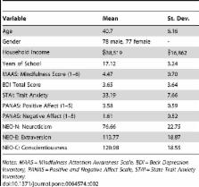
          Mindfulness, a psychological process reflecting attention and awareness to what is happening in the present moment, has been associated with increased well-being and decreased depression and anxiety in both healthy and patient populations. However, little research has explored underlying neural pathways. Recent work suggests that mindfulness (and mindfulness training interventions) may foster neuroplastic changes in cortico-limbic circuits responsible for stress and emotion regulation. Building on this work, we hypothesized that higher levels of dispositional mindfulness would be associated with decreased grey matter volume in the amgydala. In the present study, a self-report measure of dispositional mindfulness and structural MRI images were obtained from 155 healthy community adults. Volumetric analyses showed that higher dispositional mindfulness is associated with decreased grey matter volume in the right amygdala, and exploratory analyses revealed that higher dispositional mindfulness is also associated with decreased grey matter volume in the left caudate. Moreover, secondary analyses indicate that these amygdala and caudate volume associations persist after controlling for relevant demographic and individual difference factors (i.e., age, total grey matter volume, neuroticism, depression). Such volumetric differences may help explain why mindful individuals have reduced stress reactivity, and suggest new candidate structural neurobiological pathways linking mindfulness with mental and physical health outcomes.

          Related collections

          Most cited references47

          • Record: found
          • Abstract: found
          • Article: not found

          Central role of the brain in stress and adaptation: links to socioeconomic status, health, and disease.

          The brain is the key organ of stress reactivity, coping, and recovery processes. Within the brain, a distributed neural circuitry determines what is threatening and thus stressful to the individual. Instrumental brain systems of this circuitry include the hippocampus, amygdala, and areas of the prefrontal cortex. Together, these systems regulate physiological and behavioral stress processes, which can be adaptive in the short-term and maladaptive in the long-term. Importantly, such stress processes arise from bidirectional patterns of communication between the brain and the autonomic, cardiovascular, and immune systems via neural and endocrine mechanisms underpinning cognition, experience, and behavior. In one respect, these bidirectional stress mechanisms are protective in that they promote short-term adaptation (allostasis). In another respect, however, these stress mechanisms can lead to a long-term dysregulation of allostasis in that they promote maladaptive wear-and-tear on the body and brain under chronically stressful conditions (allostatic load), compromising stress resiliency and health. This review focuses specifically on the links between stress-related processes embedded within the social environment and embodied within the brain, which is viewed as the central mediator and target of allostasis and allostatic load.
            Bookmark
            • Record: found
            • Abstract: found
            • Article: not found

            Meditation experience is associated with increased cortical thickness.

            Previous research indicates that long-term meditation practice is associated with altered resting electroencephalogram patterns, suggestive of long lasting changes in brain activity. We hypothesized that meditation practice might also be associated with changes in the brain's physical structure. Magnetic resonance imaging was used to assess cortical thickness in 20 participants with extensive Insight meditation experience, which involves focused attention to internal experiences. Brain regions associated with attention, interoception and sensory processing were thicker in meditation participants than matched controls, including the prefrontal cortex and right anterior insula. Between-group differences in prefrontal cortical thickness were most pronounced in older participants, suggesting that meditation might offset age-related cortical thinning. Finally, the thickness of two regions correlated with meditation experience. These data provide the first structural evidence for experience-dependent cortical plasticity associated with meditation practice.
              Bookmark
              • Record: found
              • Abstract: found
              • Article: not found

              Amygdala, medial prefrontal cortex, and hippocampal function in PTSD.

              L Shin (2006)
              The last decade of neuroimaging research has yielded important information concerning the structure, neurochemistry, and function of the amygdala, medial prefrontal cortex, and hippocampus in posttraumatic stress disorder (PTSD). Neuroimaging research reviewed in this article reveals heightened amygdala responsivity in PTSD during symptomatic states and during the processing of trauma-unrelated affective information. Importantly, amygdala responsivity is positively associated with symptom severity in PTSD. In contrast, medial prefrontal cortex appears to be volumetrically smaller and is hyporesponsive during symptomatic states and the performance of emotional cognitive tasks in PTSD. Medial prefrontal cortex responsivity is inversely associated with PTSD symptom severity. Lastly, the reviewed research suggests diminished volumes, neuronal integrity, and functional integrity of the hippocampus in PTSD. Remaining research questions and related future directions are presented.
                Bookmark

                Author and article information

                Contributors
                Role: Editor
                Journal
                PLoS One
                PLoS ONE
                plos
                plosone
                PLoS ONE
                Public Library of Science (San Francisco, USA )
                1932-6203
                2013
                22 May 2013
                : 8
                : 5
                : e64574
                Affiliations
                [1 ]Department of Neuroscience, University of Pittsburgh, Pittsburgh, Pennsylvania, United States of America
                [2 ]Department of Psychology, Carnegie Mellon University, Pittsburgh, Pennsylvania, United States of America
                [3 ]Department of Psychology, University of Pittsburgh, Pittsburgh, Pennsylvania, United States of America
                University of Medicine & Dentistry of NJ - New Jersey Medical School, United States of America
                Author notes

                Competing Interests: The authors have declared that no competing interests exist.

                Conceived and designed the experiments: JDC AT PJG. Performed the experiments: JDC AT PJG. Analyzed the data: JDC AT PJG. Contributed reagents/materials/analysis tools: JDC AT PJG. Wrote the paper: JDC AT PJG.

                Article
                PONE-D-13-05746
                10.1371/journal.pone.0064574
                3661490
                23717632
                4ec01935-e59c-4e6b-9c79-eba31c92b537
                Copyright @ 2013

                This is an open-access article distributed under the terms of the Creative Commons Attribution License, which permits unrestricted use, distribution, and reproduction in any medium, provided the original author and source are credited.

                History
                : 7 February 2013
                : 15 April 2013
                Page count
                Pages: 7
                Funding
                This research was supported by the National Institute of Health under Grant #R01 HL089850, and the Pittsburgh Life Sciences Greenhouse Opportunity Fund. The funders had no role in study design, data collection and analysis, decision to publish, or preparation of the manuscript.
                Categories
                Research Article
                Biology
                Anatomy and Physiology
                Neurological System
                Neuroscience
                Neuroimaging
                Fmri
                Behavioral Neuroscience
                Cognitive Neuroscience
                Medicine
                Mental Health
                Psychology
                Neuropsychology
                Psychological Stress
                Social and Behavioral Sciences
                Psychology
                Neuropsychology

                Uncategorized
                Uncategorized

                Comments

                Comment on this article