19
views
0
recommends
+1 Recommend
1 collections
    0
    shares
      • Record: found
      • Abstract: found
      • Article: found
      Is Open Access

      Prediction of causative genes in inherited retinal disorder from fundus photography and autofluorescence imaging using deep learning techniques

      research-article

      Read this article at

      ScienceOpenPublisherPMC
          There is no author summary for this article yet. Authors can add summaries to their articles on ScienceOpen to make them more accessible to a non-specialist audience.

          Abstract

          Background/Aims

          To investigate the utility of a data-driven deep learning approach in patients with inherited retinal disorder (IRD) and to predict the causative genes based on fundus photography and fundus autofluorescence (FAF) imaging.

          Methods

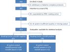
          Clinical and genetic data from 1302 subjects from 729 genetically confirmed families with IRD registered with the Japan Eye Genetics Consortium were reviewed. Three categories of genetic diagnosis were selected, based on the high prevalence of their causative genes: Stargardt disease ( ABCA4), retinitis pigmentosa ( EYS) and occult macular dystrophy ( RP1L1). Fundus photographs and FAF images were cropped in a standardised manner with a macro algorithm. Images for training/testing were selected using a randomised, fourfold cross-validation method. The application program interface was established to reach the learning accuracy of concordance (target: >80%) between the genetic diagnosis and the machine diagnosis ( ABCA4, EYS, RP1L1 and normal).

          Results

          A total of 417 images from 156 Japanese subjects were examined, including 115 genetically confirmed patients caused by the three prevalent causative genes and 41 normal subjects. The mean overall test accuracy for fundus photographs and FAF images was 88.2% and 81.3%, respectively. The mean overall sensitivity/specificity values for fundus photographs and FAF images were 88.3%/97.4% and 81.8%/95.5%, respectively.

          Conclusion

          A novel application of deep neural networks in the prediction of the causative IRD genes from fundus photographs and FAF, with a high prediction accuracy of over 80%, was highlighted. These achievements will extensively promote the quality of medical care by facilitating early diagnosis, especially by non-specialists, access to care, reducing the cost of referrals, and preventing unnecessary clinical and genetic testing.

          Related collections

          Most cited references36

          • Record: found
          • Abstract: found
          • Article: not found

          Dermatologist-level classification of skin cancer with deep neural networks

          Skin cancer, the most common human malignancy, is primarily diagnosed visually, beginning with an initial clinical screening and followed potentially by dermoscopic analysis, a biopsy and histopathological examination. Automated classification of skin lesions using images is a challenging task owing to the fine-grained variability in the appearance of skin lesions. Deep convolutional neural networks (CNNs) show potential for general and highly variable tasks across many fine-grained object categories. Here we demonstrate classification of skin lesions using a single CNN, trained end-to-end from images directly, using only pixels and disease labels as inputs. We train a CNN using a dataset of 129,450 clinical images—two orders of magnitude larger than previous datasets—consisting of 2,032 different diseases. We test its performance against 21 board-certified dermatologists on biopsy-proven clinical images with two critical binary classification use cases: keratinocyte carcinomas versus benign seborrheic keratoses; and malignant melanomas versus benign nevi. The first case represents the identification of the most common cancers, the second represents the identification of the deadliest skin cancer. The CNN achieves performance on par with all tested experts across both tasks, demonstrating an artificial intelligence capable of classifying skin cancer with a level of competence comparable to dermatologists. Outfitted with deep neural networks, mobile devices can potentially extend the reach of dermatologists outside of the clinic. It is projected that 6.3 billion smartphone subscriptions will exist by the year 2021 (ref. 13) and can therefore potentially provide low-cost universal access to vital diagnostic care.
            Bookmark
            • Record: found
            • Abstract: found
            • Article: found

            Clinically applicable deep learning for diagnosis and referral in retinal disease

              Bookmark
              • Record: found
              • Abstract: found
              • Article: found
              Is Open Access

              Pivotal trial of an autonomous AI-based diagnostic system for detection of diabetic retinopathy in primary care offices

              Artificial Intelligence (AI) has long promised to increase healthcare affordability, quality and accessibility but FDA, until recently, had never authorized an autonomous AI diagnostic system. This pivotal trial of an AI system to detect diabetic retinopathy (DR) in people with diabetes enrolled 900 subjects, with no history of DR at primary care clinics, by comparing to Wisconsin Fundus Photograph Reading Center (FPRC) widefield stereoscopic photography and macular Optical Coherence Tomography (OCT), by FPRC certified photographers, and FPRC grading of Early Treatment Diabetic Retinopathy Study Severity Scale (ETDRS) and Diabetic Macular Edema (DME). More than mild DR (mtmDR) was defined as ETDRS level 35 or higher, and/or DME, in at least one eye. AI system operators underwent a standardized training protocol before study start. Median age was 59 years (range, 22–84 years); among participants, 47.5% of participants were male; 16.1% were Hispanic, 83.3% not Hispanic; 28.6% African American and 63.4% were not; 198 (23.8%) had mtmDR. The AI system exceeded all pre-specified superiority endpoints at sensitivity of 87.2% (95% CI, 81.8–91.2%) (>85%), specificity of 90.7% (95% CI, 88.3–92.7%) (>82.5%), and imageability rate of 96.1% (95% CI, 94.6–97.3%), demonstrating AI’s ability to bring specialty-level diagnostics to primary care settings. Based on these results, FDA authorized the system for use by health care providers to detect more than mild DR and diabetic macular edema, making it, the first FDA authorized autonomous AI diagnostic system in any field of medicine, with the potential to help prevent vision loss in thousands of people with diabetes annually. ClinicalTrials.gov NCT02963441
                Bookmark

                Author and article information

                Journal
                Br J Ophthalmol
                Br J Ophthalmol
                bjophthalmol
                bjo
                The British Journal of Ophthalmology
                BMJ Publishing Group (BMA House, Tavistock Square, London, WC1H 9JR )
                0007-1161
                1468-2079
                September 2021
                20 April 2021
                : 105
                : 9
                : 1272-1279
                Affiliations
                [1 ]departmentLaboratory of Visual Physiology, Division of Vision Research , National Institute of Sensory Organs, National Hospital Organization Tokyo Medical Center , Tokyo, Japan
                [2 ]departmentDepartment of Health Policy and Management , School of Medicine, Keio University , Tokyo, Japan
                [3 ]departmentUCL Institute of Ophthalmology , UCL , London, UK
                [4 ]departmentGraduate School of Health Management , Keio University , Tokyo, Japan
                [5 ]departmentDivision of Inherited Eye Disease, Medical Retina , Moorfields Eye Hostpial , London, UK
                [6 ]departmentDivision of Molecular and Cellular Biology , National Institute of Sensory Organs, National Hospital Organization Tokyo Medical Center , Tokyo, Japan
                [7 ]departmentDepartment of Preventive Medicine and Public Health , Keio University School of Medicine , Tokyo, Japan
                [8 ]departmentSports Medicine Research Center , Keio University , Tokyo, Japan
                [9 ]departmentDivision of Vision Research , National Institute of Sensory Organs, National Hospital Organization Tokyo Medical Center , Tokyo, Japan
                Author notes
                [Correspondence to ] Dr Kaoru Fujinami, Laboratory of Visual Physiology/Ophthalmic Genetics, Tokyo Iryo Center, Meguro-ku, Tokyo, Japan; k.fujinami@ 123456ucl.ac.uk
                Author information
                http://orcid.org/0000-0001-7555-5797
                http://orcid.org/0000-0003-1782-4711
                http://orcid.org/0000-0003-4248-0033
                Article
                bjophthalmol-2020-318544
                10.1136/bjophthalmol-2020-318544
                8380883
                33879469
                c67edf25-02cf-4d67-ad4f-c9d20e743922
                © Author(s) (or their employer(s)) 2021. Re-use permitted under CC BY-NC. No commercial re-use. See rights and permissions. Published by BMJ.

                This is an open access article distributed in accordance with the Creative Commons Attribution Non Commercial (CC BY-NC 4.0) license, which permits others to distribute, remix, adapt, build upon this work non-commercially, and license their derivative works on different terms, provided the original work is properly cited, appropriate credit is given, any changes made indicated, and the use is non-commercial. See:  http://creativecommons.org/licenses/by-nc/4.0/.

                History
                : 01 December 2020
                : 12 March 2021
                : 28 March 2021
                Funding
                Funded by: FundRef http://dx.doi.org/10.13039/501100000262, Foundation Fighting Blindness;
                Award ID: CF-CL-0416-0696-UCL
                Funded by: FundRef http://dx.doi.org/10.13039/100004324, Astellas Pharma US;
                Award ID: NCT03281005
                Funded by: FundRef http://dx.doi.org/10.13039/501100001691, Japan Society for the Promotion of Science;
                Award ID: 16H06269
                Award ID: 16KK01930002
                Funded by: FundRef http://dx.doi.org/10.13039/100009619, Japan Agency for Medical Research and Development;
                Award ID: 18992608
                Funded by: National Hospital Organization;
                Award ID: H30-NHO-Sensory Organs-03
                Funded by: FundRef http://dx.doi.org/10.13039/501100003478, Ministry of Health, Labour and Welfare;
                Award ID: 18ek0109355h0001
                Categories
                Clinical Science
                1506
                Custom metadata
                unlocked

                Ophthalmology & Optometry
                retina,genetics,imaging
                Ophthalmology & Optometry
                retina, genetics, imaging

                Comments

                Comment on this article