6
views
0
recommends
+1 Recommend
0 collections
    0
    shares
      • Record: found
      • Abstract: found
      • Article: found
      Is Open Access

      Addressing Key Clinical Care and Clinical Research Needs in Severe Pediatric Traumatic Brain Injury: Perspectives From a Focused International Conference

      review-article

      Read this article at

          There is no author summary for this article yet. Authors can add summaries to their articles on ScienceOpen to make them more accessible to a non-specialist audience.

          Abstract

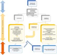
          Traumatic brain injury (TBI) is a leading cause of morbidity and mortality in children and adolescents. Survivors of severe TBI are more prone to functional deficits, resulting in poorer school performance, poor health-related quality of life (HRQoL), and increased risk of mental health problems. Critical gaps in knowledge of pathophysiological differences between children and adults concerning TBI outcomes, the paucity of pediatric trials and prognostic models and the uncertain extrapolation of adult data to pediatrics pose significant challenges and demand global efforts. Here, we explore the clinical and research unmet needs focusing on severe pediatric TBI to identify best practices in pathways of care and optimize both inpatient and outpatient management of children following TBI.

          Related collections

          Most cited references121

          • Record: found
          • Abstract: found
          • Article: not found

          Clinical characteristics and intrauterine vertical transmission potential of COVID-19 infection in nine pregnant women: a retrospective review of medical records

          Summary Background Previous studies on the pneumonia outbreak caused by the 2019 novel coronavirus disease (COVID-19) were based on information from the general population. Limited data are available for pregnant women with COVID-19 pneumonia. This study aimed to evaluate the clinical characteristics of COVID-19 in pregnancy and the intrauterine vertical transmission potential of COVID-19 infection. Methods Clinical records, laboratory results, and chest CT scans were retrospectively reviewed for nine pregnant women with laboratory-confirmed COVID-19 pneumonia (ie, with maternal throat swab samples that were positive for severe acute respiratory syndrome coronavirus 2 [SARS-CoV-2]) who were admitted to Zhongnan Hospital of Wuhan University, Wuhan, China, from Jan 20 to Jan 31, 2020. Evidence of intrauterine vertical transmission was assessed by testing for the presence of SARS-CoV-2 in amniotic fluid, cord blood, and neonatal throat swab samples. Breastmilk samples were also collected and tested from patients after the first lactation. Findings All nine patients had a caesarean section in their third trimester. Seven patients presented with a fever. Other symptoms, including cough (in four of nine patients), myalgia (in three), sore throat (in two), and malaise (in two), were also observed. Fetal distress was monitored in two cases. Five of nine patients had lymphopenia (<1·0 × 10⁹ cells per L). Three patients had increased aminotransferase concentrations. None of the patients developed severe COVID-19 pneumonia or died, as of Feb 4, 2020. Nine livebirths were recorded. No neonatal asphyxia was observed in newborn babies. All nine livebirths had a 1-min Apgar score of 8–9 and a 5-min Apgar score of 9–10. Amniotic fluid, cord blood, neonatal throat swab, and breastmilk samples from six patients were tested for SARS-CoV-2, and all samples tested negative for the virus. Interpretation The clinical characteristics of COVID-19 pneumonia in pregnant women were similar to those reported for non-pregnant adult patients who developed COVID-19 pneumonia. Findings from this small group of cases suggest that there is currently no evidence for intrauterine infection caused by vertical transmission in women who develop COVID-19 pneumonia in late pregnancy. Funding Hubei Science and Technology Plan, Wuhan University Medical Development Plan.
            Bookmark
            • Record: found
            • Abstract: found
            • Article: not found

            SARS-CoV-2 Infection in Children

            To the Editor: As of March 10, 2020, the 2019 novel coronavirus (SARS-CoV-2) has been responsible for more than 110,000 infections and 4000 deaths worldwide, but data regarding the epidemiologic characteristics and clinical features of infected children are limited. 1-3 A recent review of 72,314 cases by the Chinese Center for Disease Control and Prevention showed that less than 1% of the cases were in children younger than 10 years of age. 2 In order to determine the spectrum of disease in children, we evaluated children infected with SARS-CoV-2 and treated at the Wuhan Children’s Hospital, the only center assigned by the central government for treating infected children under 16 years of age in Wuhan. Both symptomatic and asymptomatic children with known contact with persons having confirmed or suspected SARS-CoV-2 infection were evaluated. Nasopharyngeal or throat swabs were obtained for detection of SARS-CoV-2 RNA by established methods. 4 The clinical outcomes were monitored up to March 8, 2020. Of the 1391 children assessed and tested from January 28 through February 26, 2020, a total of 171 (12.3%) were confirmed to have SARS-CoV-2 infection. Demographic data and clinical features are summarized in Table 1. (Details of the laboratory and radiologic findings are provided in the Supplementary Appendix, available with the full text of this letter at NEJM.org.) The median age of the infected children was 6.7 years. Fever was present in 41.5% of the children at any time during the illness. Other common signs and symptoms included cough and pharyngeal erythema. A total of 27 patients (15.8%) did not have any symptoms of infection or radiologic features of pneumonia. A total of 12 patients had radiologic features of pneumonia but did not have any symptoms of infection. During the course of hospitalization, 3 patients required intensive care support and invasive mechanical ventilation; all had coexisting conditions (hydronephrosis, leukemia [for which the patient was receiving maintenance chemotherapy], and intussusception). Lymphopenia (lymphocyte count, <1.2×10⁹ per liter) was present in 6 patients (3.5%). The most common radiologic finding was bilateral ground-glass opacity (32.7%). As of March 8, 2020, there was one death. A 10-month-old child with intussusception had multiorgan failure and died 4 weeks after admission. A total of 21 patients were in stable condition in the general wards, and 149 have been discharged from the hospital. This report describes a spectrum of illness from SARS-CoV-2 infection in children. In contrast with infected adults, most infected children appear to have a milder clinical course. Asymptomatic infections were not uncommon. 2 Determination of the transmission potential of these asymptomatic patients is important for guiding the development of measures to control the ongoing pandemic.
              Bookmark
              • Record: found
              • Abstract: found
              • Article: not found

              Radiation exposure from CT scans in childhood and subsequent risk of leukaemia and brain tumours: a retrospective cohort study

              Summary Background Although CT scans are very useful clinically, potential cancer risks exist from associated ionising radiation, in particular for children who are more radiosensitive than adults. We aimed to assess the excess risk of leukaemia and brain tumours after CT scans in a cohort of children and young adults. Methods In our retrospective cohort study, we included patients without previous cancer diagnoses who were first examined with CT in National Health Service (NHS) centres in England, Wales, or Scotland (Great Britain) between 1985 and 2002, when they were younger than 22 years of age. We obtained data for cancer incidence, mortality, and loss to follow-up from the NHS Central Registry from Jan 1, 1985, to Dec 31, 2008. We estimated absorbed brain and red bone marrow doses per CT scan in mGy and assessed excess incidence of leukaemia and brain tumours cancer with Poisson relative risk models. To avoid inclusion of CT scans related to cancer diagnosis, follow-up for leukaemia began 2 years after the first CT and for brain tumours 5 years after the first CT. Findings During follow-up, 74 of 178 604 patients were diagnosed with leukaemia and 135 of 176 587 patients were diagnosed with brain tumours. We noted a positive association between radiation dose from CT scans and leukaemia (excess relative risk [ERR] per mGy 0·036, 95% CI 0·005–0·120; p=0·0097) and brain tumours (0·023, 0·010–0·049; p<0·0001). Compared with patients who received a dose of less than 5 mGy, the relative risk of leukaemia for patients who received a cumulative dose of at least 30 mGy (mean dose 51·13 mGy) was 3·18 (95% CI 1·46–6·94) and the relative risk of brain cancer for patients who received a cumulative dose of 50–74 mGy (mean dose 60·42 mGy) was 2·82 (1·33–6·03). Interpretation Use of CT scans in children to deliver cumulative doses of about 50 mGy might almost triple the risk of leukaemia and doses of about 60 mGy might triple the risk of brain cancer. Because these cancers are relatively rare, the cumulative absolute risks are small: in the 10 years after the first scan for patients younger than 10 years, one excess case of leukaemia and one excess case of brain tumour per 10 000 head CT scans is estimated to occur. Nevertheless, although clinical benefits should outweigh the small absolute risks, radiation doses from CT scans ought to be kept as low as possible and alternative procedures, which do not involve ionising radiation, should be considered if appropriate. Funding US National Cancer Institute and UK Department of Health.
                Bookmark

                Author and article information

                Contributors
                Journal
                Front Pediatr
                Front Pediatr
                Front. Pediatr.
                Frontiers in Pediatrics
                Frontiers Media S.A.
                2296-2360
                18 January 2021
                2020
                : 8
                : 594425
                Affiliations
                [1] 1Pediatric Intensive Care Unit, Department of Anesthesia and Intensive Care, Papa Giovanni XXIII Hospital , Bergamo, Italy
                [2] 2Fondazione per la Ricerca dell'Ospedale di Bergamo Research Foundation, Papa Giovanni XXIII Hospital , Bergamo, Italy
                [3] 3Department of Critical Care Medicine, Safar Center for Resuscitation Research, John G Rangos Research Center, University of Pittsburgh Medical Center Children's Hospital of Pittsburgh, University of Pittsburgh School of Medicine , Pittsburgh, PA, United States
                Author notes

                Edited by: Jan Hau Lee, KK Women's and Children's Hospital, Singapore

                Reviewed by: Lee J. Martin, Johns Hopkins University, United States; Jhuma Sankar, All India Institute of Medical Sciences, India; Shu-Ling Chong, KK Women's and Children's Hospital, Singapore

                This article was submitted to Pediatric Critical Care, a section of the journal Frontiers in Pediatrics

                †The Collaborative Pediatric TBI Working Group authors are listed at the end of the article.

                Article
                10.3389/fped.2020.594425
                7849211
                33537259
                7ef7b232-6d2d-4162-8fc2-21198c8d87cc
                Copyright © 2021 Nacoti, Fazzi, Biroli, Zangari, Barbui, Kochanek and the Collaborative Pediatric TBI Working Group.

                This is an open-access article distributed under the terms of the Creative Commons Attribution License (CC BY). The use, distribution or reproduction in other forums is permitted, provided the original author(s) and the copyright owner(s) are credited and that the original publication in this journal is cited, in accordance with accepted academic practice. No use, distribution or reproduction is permitted which does not comply with these terms.

                History
                : 13 August 2020
                : 06 November 2020
                Page count
                Figures: 2, Tables: 2, Equations: 0, References: 125, Pages: 17, Words: 14444
                Categories
                Pediatrics
                Review

                traumatic brain injury,pediatric,intracranial pressure,outcomes,prognosis

                Comments

                Comment on this article