2
views
0
recommends
+1 Recommend
0 collections
    0
    shares
      • Record: found
      • Abstract: found
      • Article: found
      Is Open Access

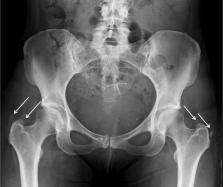
      Bilateral gluteus medius and minimus calcific tendonitis in a patient with previous bilateral calcific tendonitis of the shoulder: A case report

      case-report
      , MBBS, iBSc, FRCR a , * , , MBBS, MRCGP b , , MBBS, PhD, FRCP a , , MBBS, D'Orth, FCPS, MS, MCh.Orth, FRCS Orth c , , MBBS, MRCS, MS, FRCR a
      Radiology Case Reports
      Elsevier
      Calcific, Tendonitis, Musculoskeletal, Gluteus, Tendinopathy

      Read this article at

          There is no author summary for this article yet. Authors can add summaries to their articles on ScienceOpen to make them more accessible to a non-specialist audience.

          Abstract

          Calcific tendonitis of the gluteus medius and minimus tendons is a rare complication of hydroxyapatite deposition disease, with bilateral involvement even more so. Although patients can be asymptomatic, there is often an acute-on-chronic presentation of pain. We present a case of bilateral calcific tendonitis of the gluteus medius and minimus tendons on a background of previous bilateral rotator cuff calcific tendonitis in a middle-aged woman. This patient's long-standing history of multi-focal involvement required a multidisciplinary approach between orthopedics, rheumatology, and musculoskeletal radiology for optimal management, requiring different treatment options for different affected sites.

          Related collections

          Most cited references18

          • Record: found
          • Abstract: found
          • Article: not found

          Calcific Tendinopathy of the Rotator Cuff: Pathogenesis, Diagnosis, and Management.

          Calcific tendinopathy, or calcifying tendinitis, is a disease characterized by multifocal, cell-mediated calcification of living tissue. After spontaneous disappearance of the calcific deposits or, less frequently, surgical removal, the tendon reconstitutes itself. Attention to the clinical presentation and the radiologic, morphologic, and gross characteristics of the calcium deposit will facilitate differentiation between the formative phase and the resorptive phase, which is of paramount importance in the management of this disease. Should conservative treatment fail, surgical removal may be indicated during the formative phase, but only under exceptional circumstances during the resorptive phase. Aspiration and lavage of the deposit should be performed only during the latter phase.
            Bookmark
            • Record: found
            • Abstract: found
            • Article: not found

            Gluteal Tendinopathy: A Review of Mechanisms, Assessment and Management

            Tendinopathy of the gluteus medius and gluteus minimus tendons is now recognized as a primary local source of lateral hip pain. The condition mostly occurs in mid-life both in athletes and in subjects who do not regularly exercise. Females are afflicted more than males. This condition interferes with sleep (side lying) and common weight-bearing tasks, which makes it a debilitating musculoskeletal condition with a significant impact. Mechanical loading drives the biological processes within a tendon and determines its structural form and load-bearing capacity. The combination of excessive compression and high tensile loads within tendons are thought to be most damaging. The available evidence suggests that joint position (particularly excessive hip adduction), together with muscle and bone elements, are key factors in gluteal tendinopathy. These factors provide a basis for a clinical reasoning process in the assessment and management of a patient presenting with localized lateral hip pain from gluteal tendinopathy. Currently, there is a lack of consensus as to which clinical examination tests provide best diagnostic utility. On the basis of the few diagnostic utility studies and the current understanding of the pathomechanics of gluteal tendinopathy, we propose that a battery of clinical tests utilizing a combination of provocative compressive and tensile loads is currently best practice in its assessment. Management of this condition commonly involves corticosteroid injection, exercise or shock wave therapy, with surgery reserved for recalcitrant cases. There is a dearth of evidence for any treatments, so the approach we recommend involves managing the load on the tendons through exercise and education on the underlying pathomechanics.
              Bookmark
              • Record: found
              • Abstract: found
              • Article: not found

              Calcific tendinitis: a pictorial review.

              Calcific tendinitis is caused by the pathologic deposition of calcium hydroxyapatite crystals in tendons and is a common cause of joint pain. The disease typically affects the shoulder and hip, with characteristic imaging findings; however, any joint can be involved. Occasionally, calcific tendinitis can mimic aggressive disorders, such as infection and neoplasm, especially on magnetic resonance imaging. Radiologists should be familiar with the imaging findings to distinguish calcific tendinitis from more aggressive processes. Image-guided percutaneous needle aspiration and steroid injection of calcific tendinitis are useful techniques performed by the radiologist for the treatment of symptomatic cases. Familiarity with these procedures and their imaging appearance is an important aspect in the management of this common disease.
                Bookmark

                Author and article information

                Contributors
                Journal
                Radiol Case Rep
                Radiol Case Rep
                Radiology Case Reports
                Elsevier
                1930-0433
                26 August 2021
                November 2021
                26 August 2021
                : 16
                : 11
                : 3222-3225
                Affiliations
                [a ]London North West University Healthcare NHS Trust, London, England
                [b ]Practice Plus Group, London, England
                [c ]BMI The Clementine Churchill Hospital, London, England
                Author notes
                [* ] Corresponding author. mostafa.ellatif@ 123456nhs.net
                Article
                S1930-0433(21)00546-X
                10.1016/j.radcr.2021.07.074
                8405919
                50a0f6ce-460c-4224-bdeb-a894d143c99a
                © 2021 The Authors. Published by Elsevier Inc. on behalf of University of Washington.

                This is an open access article under the CC BY-NC-ND license (http://creativecommons.org/licenses/by-nc-nd/4.0/).

                History
                : 22 July 2021
                : 25 July 2021
                : 26 July 2021
                Categories
                Case Report

                calcific,tendonitis,musculoskeletal,gluteus,tendinopathy
                calcific, tendonitis, musculoskeletal, gluteus, tendinopathy

                Comments

                Comment on this article