55
views
0
recommends
+1 Recommend
0 collections
    0
    shares
      • Record: found
      • Abstract: found
      • Article: found
      Is Open Access

      Dynamic blastomere behaviour reflects human embryo ploidy by the four-cell stage

      research-article

      Read this article at

          There is no author summary for this article yet. Authors can add summaries to their articles on ScienceOpen to make them more accessible to a non-specialist audience.

          Abstract

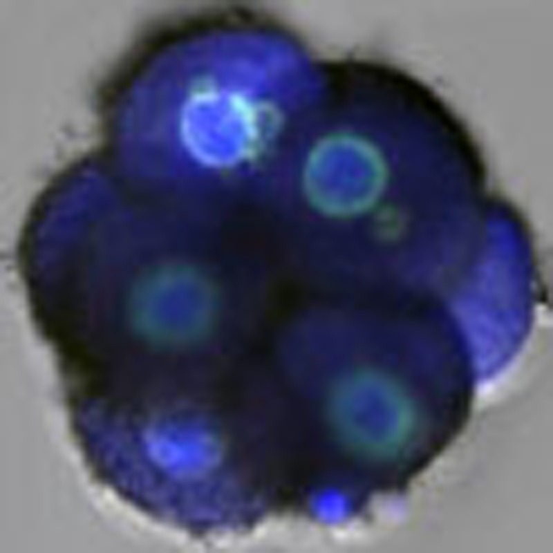
          Previous studies have demonstrated that aneuploidy in human embryos is surprisingly frequent with 50–80% of cleavage-stage human embryos carrying an abnormal chromosome number. Here we combine non-invasive time-lapse imaging with karyotypic reconstruction of all blastomeres in four-cell human embryos to address the hypothesis that blastomere behaviour may reflect ploidy during the first two cleavage divisions. We demonstrate that precise cell cycle parameter timing is observed in all euploid embryos to the four-cell stage, whereas only 30% of aneuploid embryos exhibit parameter values within normal timing windows. Further, we observe that the generation of human embryonic aneuploidy is complex with contribution from chromosome-containing fragments/micronuclei that frequently emerge and may persist or become reabsorbed during interphase. These findings suggest that cell cycle and fragmentation parameters of individual blastomeres are diagnostic of ploidy, amenable to automated tracking algorithms, and likely of clinical relevance in reducing transfer of embryos prone to miscarriage.

          Abstract

          Abnormal human embryo development is implicated in the embryo arrest observed during in vitro fertilization. Chavez and colleagues perform time-lapse imaging on human embryos and find that chromosomally abnormal embryos exhibit diverse cell cycle parameters that may contribute to arrest.

          Related collections

          Most cited references48

          • Record: found
          • Abstract: found
          • Article: not found

          Chromosome instability is common in human cleavage-stage embryos.

          Chromosome instability is a hallmark of tumorigenesis. This study establishes that chromosome instability is also common during early human embryogenesis. A new array-based method allowed screening of genome-wide copy number and loss of heterozygosity in single cells. This revealed not only mosaicism for whole-chromosome aneuploidies and uniparental disomies in most cleavage-stage embryos but also frequent segmental deletions, duplications and amplifications that were reciprocal in sister blastomeres, implying the occurrence of breakage-fusion-bridge cycles. This explains the low human fecundity and identifies post-zygotic chromosome instability as a leading cause of constitutional chromosomal disorders.
            Bookmark
            • Record: found
            • Abstract: found
            • Article: not found

            Non-invasive imaging of human embryos before embryonic genome activation predicts development to the blastocyst stage.

            We report studies of preimplantation human embryo development that correlate time-lapse image analysis and gene expression profiling. By examining a large set of zygotes from in vitro fertilization (IVF), we find that success in progression to the blastocyst stage can be predicted with >93% sensitivity and specificity by measuring three dynamic, noninvasive imaging parameters by day 2 after fertilization, before embryonic genome activation (EGA). These parameters can be reliably monitored by automated image analysis, confirming that successful development follows a set of carefully orchestrated and predictable events. Moreover, we show that imaging phenotypes reflect molecular programs of the embryo and of individual blastomeres. Single-cell gene expression analysis reveals that blastomeres develop cell autonomously, with some cells advancing to EGA and others arresting. These studies indicate that success and failure in human embryo development is largely determined before EGA. Our methods and algorithms may provide an approach for early diagnosis of embryo potential in assisted reproduction.
              Bookmark
              • Record: found
              • Abstract: not found
              • Article: not found

              Genetic control of the cell division cycle in yeast.

                Bookmark

                Author and article information

                Journal
                Nat Commun
                Nat Commun
                Nature Communications
                Nature Pub. Group
                2041-1723
                04 December 2012
                : 3
                : 1251
                Affiliations
                [1 ]Institute for Stem Cell Biology and Regenerative Medicine, Stanford University School of Medicine , Stanford, California 94305, USA
                [2 ]Department of Obstetrics and Gynecology, Stanford University School of Medicine , Stanford, California 94305, USA
                [3 ]Auxogyn, Inc. , Menlo Park, California 94025, USA
                [4 ]Reprogenetics , Livingston, New Jersey 07039, USA
                Author notes
                Article
                ncomms2249
                10.1038/ncomms2249
                3535341
                23212380
                559334b4-bb7e-4a8c-97d1-57b99562a68a
                Copyright © 2012, Nature Publishing Group, a division of Macmillan Publishers Limited. All Rights Reserved.

                This work is licensed under a Creative Commons Attribution-NonCommercial-Share Alike 3.0 Unported License. To view a copy of this license, visit http://creativecommons.org/licenses/by-nc-sa/3.0/

                History
                : 21 February 2012
                : 01 November 2012
                Categories
                Article

                Uncategorized
                Uncategorized

                Comments

                Comment on this article