2
views
0
recommends
+1 Recommend
0 collections
    0
    shares
      • Record: found
      • Abstract: found
      • Article: found
      Is Open Access

      Inhibition of TRPV1 by SHP-1 in nociceptive primary sensory neurons is critical in PD-L1 analgesia

      research-article
      1 , 1 , 1 , 1 , 1 , 2 ,
      JCI Insight
      American Society for Clinical Investigation
      Neuroscience, Pain, Signal transduction

      Read this article at

          There is no author summary for this article yet. Authors can add summaries to their articles on ScienceOpen to make them more accessible to a non-specialist audience.

          Abstract

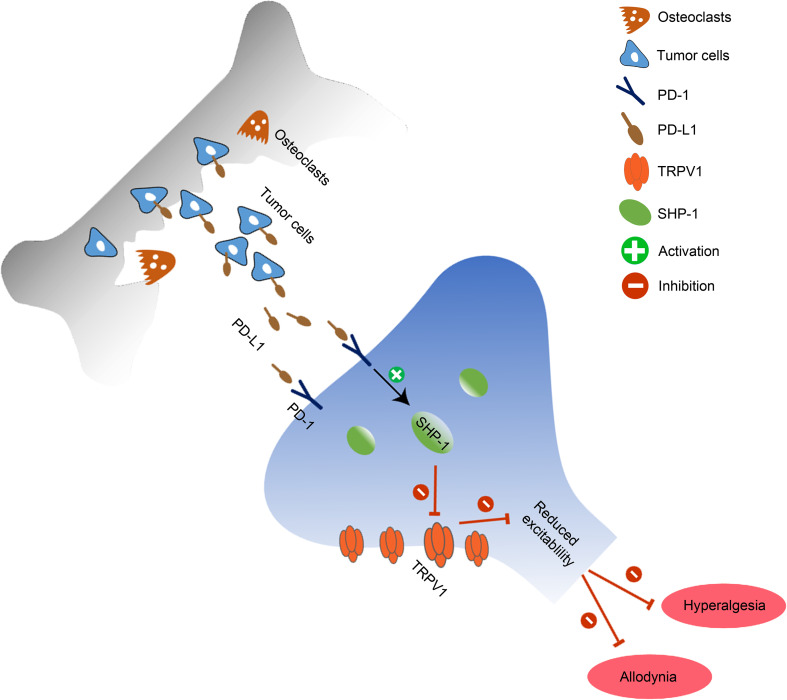
          Recently programmed death-ligand 1 (PD-L1) receptor PD-1 was found in dorsal root ganglion (DRG) neurons, and PD-L1 activates PD-1 to inhibit inflammatory and neuropathic pain by modulating neuronal excitability. However, the downstream signaling of PD-1 in sensory neurons remains unclear. Here, we show that PD-L1 activated Src homology 2 domain-containing tyrosine phosphatase-1 (SHP-1) to downregulate transient receptor potential vanilloid 1 (TRPV1) in DRG neurons and inhibit bone cancer pain in mice. Local injection of PD-L1 produced analgesia. PD-1 in DRG neurons colocalized with TRPV1 and SHP-1. PD-L1 induced the phosphorylation of SHP-1 in DRG TRPV1 neurons and inhibited TRPV1 currents. Loss of TRPV1 in mice abolished bone cancer–induced thermal hyperalgesia and PD-L1 analgesia. Conditioned deletion of SHP-1 in Na V1.8 + neurons aggravated bone cancer pain and diminished the inhibition of PD-L1 on TRPV1 currents and pain. Together, our findings suggest that PD-L1/PD-1 signaling suppresses bone cancer pain via inhibition of TRPV1 activity. Our results also suggest that SHP-1 in sensory neurons is an endogenous pain inhibitor and delays the development of bone cancer pain via suppressing TRPV1 function.

          Abstract

          Abstract

          PD-L1/PD-1 signaling suppresses TRPV1 activity and alleviates pain-like behaviors via phosphorylation of SHP-1 in nociceptive primary sensory neurons in a mouse bone cancer model.

          Related collections

          Most cited references79

          • Record: found
          • Abstract: found
          • Article: not found

          Safety and Activity of Anti–PD-L1 Antibody in Patients with Advanced Cancer

          Programmed death 1 (PD-1) protein, a T-cell coinhibitory receptor, and one of its ligands, PD-L1, play a pivotal role in the ability of tumor cells to evade the host's immune system. Blockade of interactions between PD-1 and PD-L1 enhances immune function in vitro and mediates antitumor activity in preclinical models. In this multicenter phase 1 trial, we administered intravenous anti-PD-L1 antibody (at escalating doses ranging from 0.3 to 10 mg per kilogram of body weight) to patients with selected advanced cancers. Anti-PD-L1 antibody was administered every 14 days in 6-week cycles for up to 16 cycles or until the patient had a complete response or confirmed disease progression. As of February 24, 2012, a total of 207 patients--75 with non-small-cell lung cancer, 55 with melanoma, 18 with colorectal cancer, 17 with renal-cell cancer, 17 with ovarian cancer, 14 with pancreatic cancer, 7 with gastric cancer, and 4 with breast cancer--had received anti-PD-L1 antibody. The median duration of therapy was 12 weeks (range, 2 to 111). Grade 3 or 4 toxic effects that investigators considered to be related to treatment occurred in 9% of patients. Among patients with a response that could be evaluated, an objective response (a complete or partial response) was observed in 9 of 52 patients with melanoma, 2 of 17 with renal-cell cancer, 5 of 49 with non-small-cell lung cancer, and 1 of 17 with ovarian cancer. Responses lasted for 1 year or more in 8 of 16 patients with at least 1 year of follow-up. Antibody-mediated blockade of PD-L1 induced durable tumor regression (objective response rate of 6 to 17%) and prolonged stabilization of disease (rates of 12 to 41% at 24 weeks) in patients with advanced cancers, including non-small-cell lung cancer, melanoma, and renal-cell cancer. (Funded by Bristol-Myers Squibb and others; ClinicalTrials.gov number, NCT00729664.).
            Bookmark
            • Record: found
            • Abstract: found
            • Article: not found

            PD-1 and its ligands in tolerance and immunity.

            Programmed death 1 (PD-1) and its ligands, PD-L1 and PD-L2, deliver inhibitory signals that regulate the balance between T cell activation, tolerance, and immunopathology. Immune responses to foreign and self-antigens require specific and balanced responses to clear pathogens and tumors and yet maintain tolerance. Induction and maintenance of T cell tolerance requires PD-1, and its ligand PD-L1 on nonhematopoietic cells can limit effector T cell responses and protect tissues from immune-mediated tissue damage. The PD-1:PD-L pathway also has been usurped by microorganisms and tumors to attenuate antimicrobial or tumor immunity and facilitate chronic infection and tumor survival. The identification of B7-1 as an additional binding partner for PD-L1, together with the discovery of an inhibitory bidirectional interaction between PD-L1 and B7-1, reveals new ways the B7:CD28 family regulates T cell activation and tolerance. In this review, we discuss current understanding of the immunoregulatory functions of PD-1 and its ligands and their therapeutic potential.
              Bookmark
              • Record: found
              • Abstract: found
              • Article: found
              Is Open Access

              Molecular Architecture of the Mouse Nervous System

              Summary The mammalian nervous system executes complex behaviors controlled by specialized, precisely positioned, and interacting cell types. Here, we used RNA sequencing of half a million single cells to create a detailed census of cell types in the mouse nervous system. We mapped cell types spatially and derived a hierarchical, data-driven taxonomy. Neurons were the most diverse and were grouped by developmental anatomical units and by the expression of neurotransmitters and neuropeptides. Neuronal diversity was driven by genes encoding cell identity, synaptic connectivity, neurotransmission, and membrane conductance. We discovered seven distinct, regionally restricted astrocyte types that obeyed developmental boundaries and correlated with the spatial distribution of key glutamate and glycine neurotransmitters. In contrast, oligodendrocytes showed a loss of regional identity followed by a secondary diversification. The resource presented here lays a solid foundation for understanding the molecular architecture of the mammalian nervous system and enables genetic manipulation of specific cell types.
                Bookmark

                Author and article information

                Contributors
                Journal
                JCI Insight
                JCI Insight
                JCI Insight
                JCI Insight
                American Society for Clinical Investigation
                2379-3708
                15 October 2020
                15 October 2020
                15 October 2020
                : 5
                : 20
                : e137386
                Affiliations
                [1 ]State Key Laboratory of Medical Neurobiology and MOE Frontiers Center for Brain Science, Department of Translational Neuroscience, Jing’an District Centre Hospital of Shanghai, Institutes of Brain Science, Fudan University, Shanghai, China.
                [2 ]Institutes of Integrative Medicine, Fudan University, Shanghai, China.
                Author notes
                Address correspondence to: Yu-Qiu Zhang, Institutes of Brain Science, Fudan University, 1202 Mingdao Building, 131 Dong An Road, Shanghai 200032, China. Phone: 86.021.54237635; Email: yuqiuzhang@ 123456fudan.edu.cn .

                Authorship note: BLL and QLC contributed equally to this work as co–first authors.

                Author information
                http://orcid.org/0000-0002-9646-7531
                http://orcid.org/0000-0001-6623-9629
                Article
                137386
                10.1172/jci.insight.137386
                7605531
                32960817
                a906f0d2-45c7-4f96-8d55-68d41ff10fa4
                © 2020 Liu et al.

                This work is licensed under the Creative Commons Attribution 4.0 International License. To view a copy of this license, visit http://creativecommons.org/licenses/by/4.0/.

                History
                : 18 February 2020
                : 10 September 2020
                Funding
                Funded by: National Natural Science Foundation of China
                Award ID: 31930042,31771164
                Funded by: National Key R&D Program of China
                Award ID: 2017YFB0403803
                Funded by: Shanghai Municipal Science and Technology Major Project
                Award ID: 2018SHZDZX01
                Categories
                Research Article

                neuroscience,pain,signal transduction
                neuroscience, pain, signal transduction

                Comments

                Comment on this article