7
views
0
recommends
+1 Recommend
0 collections
    0
    shares
      • Record: found
      • Abstract: found
      • Article: found
      Is Open Access

      Influence of rebubbling on anterior segment parameters and refractive outcomes in eyes with DMEK for Fuchs endothelial dystrophy

      research-article

      Read this article at

          There is no author summary for this article yet. Authors can add summaries to their articles on ScienceOpen to make them more accessible to a non-specialist audience.

          Abstract

          Purpose

          To evaluate the potential impact of rebubbling on the anterior segment parameters and refractive outcomes in patients with graft detachment following uneventful DMEK for Fuchs endothelial dystrophy (FED).

          Methods

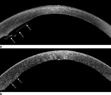
          Retrospective institutional cohort study of comparing 34 eyes of 31 patients with rebubbling for graft detachment following Descemet membrane endothelial keratoplasty (DMEK) to 33 eyes of 28 patients with uneventful DMEK. Main outcome parameters were various corneal parameters obtained by Scheimpflug imaging, refractive outcome, corrected distance visual acuity (CDVA), and endothelial cell density (ECD).

          Results

          Anterior and posterior corneal astigmatism, corneal densitometry, central corneal thickness, and anterior chamber depth and volume showed no significant differences. Preoperative distribution of astigmatism axis orientations showed a high proportion of anterior corneal with-the-rule astigmatism (71%) in eyes requiring rebubbling. Mean postoperative cylinder in the rebubbling group (1.21 ± 0.85 D) was significantly higher compared to the controls ( p = 0.04), while differences in spherical equivalent (SE) were insignificant ( p = 0.24). Postoperative CDVA was 0.11 ± 0.11 in the control group compared to 0.21 ± 0.17 in the rebubbling group ( p = 0.03). Eyes with subsequent rebubbling demonstrated a significantly higher endothelial cell loss (56% versus 37%) ( p < 0.001).

          Conclusion

          Apart from higher cylinder values, refractive outcome and corneal parameters assessed by Scheimpflug imaging were comparable in eyes with rebubbling and controls. However, a reduced visual acuity and an increased endothelial cell loss should be taken into consideration prior to rebubbling especially in eyes with circumscribed graft detachment.

          Related collections

          Most cited references23

          • Record: found
          • Abstract: found
          • Article: not found

          Descemet membrane endothelial keratoplasty (DMEK).

          To describe Descemet membrane endothelial keratoplasty (DMEK) with organ cultured Descemet membrane (DM) in a human cadaver eye model and a patient with Fuchs endothelial dystrophy. In 10 human cadaver eyes and 1 patient eye, a 3.5-mm clear corneal tunnel incision was made. The anterior chamber was filled with air, and the DM was stripped off from the posterior stroma. From organ-cultured donor corneo-scleral rims, 9.0-mm-diameter "DM rolls" were harvested. Each donor DM roll was inserted into a recipient anterior chamber, positioned onto the posterior stroma, and kept in position by completely filling the anterior chamber with air for 30 minutes. In all recipient eyes, the donor DM maintained its position after a 30-minute air-fill of the anterior chamber followed by an air-liquid exchange. In the patient's eye, 1 week after transplantation, best-corrected visual acuity was 1.0 (20/20) with the patient's preoperative refraction, and the endothelial cell density averaged 2350 cells/mm. DMEK may provide quick visual rehabilitation in the treatment of corneal endothelial disorders by transplantation of an organ-cultured DM transplanted through a clear corneal tunnel incision. DMEK may be a highly accessible procedure to corneal surgeons, because donor DM sheets can be prepared from preserved corneo-scleral rims.
            Bookmark
            • Record: found
            • Abstract: found
            • Article: not found

            Normative values for corneal densitometry analysis by Scheimpflug optical assessment.

            To describe the normative data for corneal Scheimpflug densitometry based on a cohort of normal participants. A total of 445 healthy participants were recruited for assessment (794 eyes). Left and right eyes were considered separately. All participants were assessed using the corneal densitometry analysis add-on to the standard software of the Oculus Pentacam. Densitometry measurements were obtained and expressed in standardized grayscale units (GSU). All participants were Caucasian; 42% were male and 58% were female. The mean age was 48.0 ± 15.3 years (range, 20.2-84.2 years). Mean corneal densitometry over the 12-mm-diameter area was 19.74 ± 3.89 GSU. When divided by radial zone, densitometry values were lowest in the central zone (16.76 ± 1.87 GSU) and highest in the periphery (27.36 ± 7.47 GSU). There was no difference between central zone and the surrounding 2- to 6-mm annulus (P > 0.05), though the 6- to 10-mm and the 10- to 12-mm zones displayed higher densitometry values (P < 0.001). When divided by depth, the anterior layer displayed the highest densitometry reading of 25.81 ± 5.14 GSU, which was significantly higher than that of both the central (P < 0.001) and the posterior layers (P < 0.001). Changes in corneal densitometry were correlated with age, though not within the central 6-mm-diameter ring. No sex difference was seen within the cohort. This add-on to the standard imaging software allows rapid and objective assessment of the corneal densitometry. We provide normative data that may be used as a reference facilitating research and complementing clinical examination.
              Bookmark
              • Record: found
              • Abstract: found
              • Article: not found

              Risk of corneal transplant rejection significantly reduced with Descemet's membrane endothelial keratoplasty.

              To evaluate the relative risk of immunologic rejection episode in patients who underwent Descemet's membrane endothelial keratoplasty (DMEK), Descemet's stripping endothelial keratoplasty (DSEK), and penetrating keratoplasty (PK). Comparative case series. One hundred forty-one eyes treated with DMEK at Price Vision Group, Indianapolis, Indiana. The patients in the DMEK group were compared retrospectively with cohorts of DSEK (n = 598) and PK (n = 30) patients treated at the same center, with similar demographics, follow-up duration, and indications for surgery. The postoperative steroid regimen and rejection criteria were identical in the 3 groups. Kaplan-Meier survival analysis, which takes varying length of follow-up into consideration, was performed to determine the cumulative probability of a rejection episode 1 and 2 years after surgery. Proportional hazards analysis was used to determine the relative risk of rejection episodes between the 3 groups. P<0.05 was considered significant and calculated using the log-rank test. Rejection-free survival and cumulative probability of a rejection episode. The mean recipient age was 66 years (56% females and 94% Caucasian) and median follow-up duration was 13 months (range, 3-40) in the DMEK group. Fuchs' dystrophy was the most common indication for surgery (n = 127; 90%) followed by pseudophakic bullous keratopathy (n = 4; 4%) and regrafts (n = 9; 6.4%). Only 1 patient (0.7%) had a documented rejection episode in the DMEK group compared with 54 (9%) in the DSEK and 5 (17%) in the PK group. The Kaplan-Meier cumulative probability of a rejection episode at 1 and 2 years was 1% and 1%, respectively, for DMEK; 8% and 12%, respectively, for DSEK; and 14% and 18%, respectively, for PK. This was a highly significant difference (P = 0.004). The DMEK eyes had a 15 times lesser risk of experiencing a rejection episode than DSEK eyes (95% confidence limit [CL], 2.0-111; P = 0.008) and 20 times lower risk than PK eyes (95% CL, 2.4-166; P = 0.006). Patients undergoing DMEK had a significantly reduced risk of experiencing a rejection episode within 2 years after surgery compared with DSEK and PK performed for similar indications using the same corticosteroid regimen. Copyright © 2012 American Academy of Ophthalmology. Published by Elsevier Inc. All rights reserved.
                Bookmark

                Author and article information

                Contributors
                kohnen@em.uni-frankfurt.de
                Journal
                Graefes Arch Clin Exp Ophthalmol
                Graefes Arch Clin Exp Ophthalmol
                Graefe's Archive for Clinical and Experimental Ophthalmology
                Springer Berlin Heidelberg (Berlin/Heidelberg )
                0721-832X
                1435-702X
                6 April 2021
                6 April 2021
                2021
                : 259
                : 10
                : 3175-3183
                Affiliations
                GRID grid.7839.5, ISNI 0000 0004 1936 9721, Department of Ophthalmology, , Goethe-University, ; Theodor-Stern-Kai 7, 60590 Frankfurt am Main, Germany
                Author information
                http://orcid.org/0000-0002-6933-9585
                Article
                5114
                10.1007/s00417-021-05114-2
                8478761
                33825029
                b95b35af-593d-4599-ba2b-cdea6d677ccf
                © The Author(s) 2021

                Open Access This article is licensed under a Creative Commons Attribution 4.0 International License, which permits use, sharing, adaptation, distribution and reproduction in any medium or format, as long as you give appropriate credit to the original author(s) and the source, provide a link to the Creative Commons licence, and indicate if changes were made. The images or other third party material in this article are included in the article's Creative Commons licence, unless indicated otherwise in a credit line to the material. If material is not included in the article's Creative Commons licence and your intended use is not permitted by statutory regulation or exceeds the permitted use, you will need to obtain permission directly from the copyright holder. To view a copy of this licence, visit http://creativecommons.org/licenses/by/4.0/.

                History
                : 29 October 2020
                : 4 January 2021
                : 8 February 2021
                Funding
                Funded by: Johann Wolfgang Goethe-Universität, Frankfurt am Main (1022)
                Categories
                Refractive Surgery
                Custom metadata
                © Springer-Verlag GmbH Germany, part of Springer Nature 2021

                Ophthalmology & Optometry
                rebubbling,graft detachment,descemet membrane endothelial keratoplasty (dmek),fuchs endothelial dystrophy (fed),endothelial cell density (ecd)

                Comments

                Comment on this article