3
views
0
recommends
+1 Recommend
0 collections
    0
    shares
      • Record: found
      • Abstract: found
      • Article: found
      Is Open Access

      Disruption of white matter integrity and its relationship with cognitive function in non-severe traumatic brain injury

      research-article
      1 , 1 , 2 , * , , 1 , 3 , 2 , 4 , 5 , 2 , 4 , 2 , 4 , 5 , 2 , 4 , 2 , 5 , 2 , 4 , 5 , 2 , 4 , 5 , 2 , 4 , 5 , 2 , 4 , 5 , 2 , 4 , 5 , 2 , 4 , 5 , 2 , 4 , 5 , 2 , 4 , 5 , 2 , 4 , 5 , 2 , 4 , 5 , 6 , 7 , 8 , 9 , 10 , 2 , 11 , 2 , 11 , 12 , 13 , 14 , 13 , 14 , 15 , 16 , 17 , 18 , 19 , 20 , 20 , 2 , 4 , 5
      Frontiers in Neurology
      Frontiers Media S.A.
      traumatic brain injury, diffusion MRI, fractional anisotropy, neuropsychological test, tract-based spatial statistic

      Read this article at

          There is no author summary for this article yet. Authors can add summaries to their articles on ScienceOpen to make them more accessible to a non-specialist audience.

          Abstract

          Background

          Impairment in cognitive function is a recognized outcome of traumatic brain injury (TBI). However, the degree of impairment has variable relationship with TBI severity and time post injury. The underlying pathology is often due to diffuse axonal injury that has been found even in mild TBI. In this study, we examine the state of white matter putative connectivity in patients with non-severe TBI in the subacute phase, i.e., within 10 weeks of injury and determine its relationship with neuropsychological scores.

          Methods

          We conducted a case-control prospective study involving 11 male adult patients with non-severe TBI and an age-matched control group of 11 adult male volunteers. Diffusion MRI scanning and neuropsychological tests were administered within 10 weeks post injury. The difference in fractional anisotropy (FA) values between the patient and control groups was examined using tract-based spatial statistics. The FA values that were significantly different between patients and controls were then correlated with neuropsychological tests in the patient group.

          Results

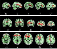
          Several clusters with peak voxels of significant FA reductions ( p < 0.05) in the white matter skeleton were seen in patients compared to the control group. These clusters were located in the superior fronto-occipital fasciculus, superior longitudinal fasciculus, uncinate fasciculus, and cingulum, as well as white matter fibers in the area of genu of corpus callosum, anterior corona radiata, superior corona radiata, anterior thalamic radiation and part of inferior frontal gyrus. Mean global FA magnitude correlated significantly with MAVLT immediate recall scores while matrix reasoning scores correlated positively with FA values in the area of right superior fronto-occipital fasciculus and left anterior corona radiata.

          Conclusion

          The non-severe TBI patients had abnormally reduced FA values in multiple regions compared to controls that correlated with several measures of executive function during the sub-acute phase of TBI.

          Related collections

          Most cited references70

          • Record: found
          • Abstract: not found
          • Article: not found

          The assessment and analysis of handedness: The Edinburgh inventory

            Bookmark
            • Record: found
            • Abstract: found
            • Article: not found

            FSL.

            FSL (the FMRIB Software Library) is a comprehensive library of analysis tools for functional, structural and diffusion MRI brain imaging data, written mainly by members of the Analysis Group, FMRIB, Oxford. For this NeuroImage special issue on "20 years of fMRI" we have been asked to write about the history, developments and current status of FSL. We also include some descriptions of parts of FSL that are not well covered in the existing literature. We hope that some of this content might be of interest to users of FSL, and also maybe to new research groups considering creating, releasing and supporting new software packages for brain image analysis. Copyright © 2011 Elsevier Inc. All rights reserved.
              Bookmark
              • Record: found
              • Abstract: found
              • Article: not found

              Fast robust automated brain extraction.

              An automated method for segmenting magnetic resonance head images into brain and non-brain has been developed. It is very robust and accurate and has been tested on thousands of data sets from a wide variety of scanners and taken with a wide variety of MR sequences. The method, Brain Extraction Tool (BET), uses a deformable model that evolves to fit the brain's surface by the application of a set of locally adaptive model forces. The method is very fast and requires no preregistration or other pre-processing before being applied. We describe the new method and give examples of results and the results of extensive quantitative testing against "gold-standard" hand segmentations, and two other popular automated methods. Copyright 2002 Wiley-Liss, Inc.
                Bookmark

                Author and article information

                Contributors
                URI : http://loop.frontiersin.org/people/1968897/overview
                URI : http://loop.frontiersin.org/people/1613739/overview
                URI : http://loop.frontiersin.org/people/1692400/overview
                URI : http://loop.frontiersin.org/people/226719/overview
                URI : http://loop.frontiersin.org/people/491506/overview
                URI : http://loop.frontiersin.org/people/671153/overview
                URI : http://loop.frontiersin.org/people/1608601/overview
                URI : http://loop.frontiersin.org/people/198149/overview
                URI : http://loop.frontiersin.org/people/1741767/overview
                URI : http://loop.frontiersin.org/people/490417/overview
                URI : http://loop.frontiersin.org/people/1613784/overview
                URI : http://loop.frontiersin.org/people/1529472/overview
                URI : http://loop.frontiersin.org/people/1607889/overview
                URI : http://loop.frontiersin.org/people/1128373/overview
                URI : http://loop.frontiersin.org/people/1613767/overview
                URI : http://loop.frontiersin.org/people/712358/overview
                URI : http://loop.frontiersin.org/people/54366/overview
                URI : http://loop.frontiersin.org/people/125027/overview
                URI : http://loop.frontiersin.org/people/1614520/overview
                URI : http://loop.frontiersin.org/people/1664888/overview
                URI : http://loop.frontiersin.org/people/44636/overview
                Journal
                Front Neurol
                Front Neurol
                Front. Neurol.
                Frontiers in Neurology
                Frontiers Media S.A.
                1664-2295
                11 October 2022
                2022
                : 13
                : 1011304
                Affiliations
                [1] 1Department of Physiology, School of Medical Sciences, Universiti Sains Malaysia , Kota Bharu, Malaysia
                [2] 2Brain and Behaviour Cluster, School of Medical Sciences, Universiti Sains Malaysia , Kota Bharu, Malaysia
                [3] 3Faculty of Science and Technology, Universiti Sains Islam Malaysia , Nilai, Malaysia
                [4] 4Department of Neurosciences, School of Medical Sciences, Universiti Sains Malaysia , Kota Bharu, Malaysia
                [5] 5Hospital Universiti Sains Malaysia, Universiti Sains Malaysia , Kota Bharu, Malaysia
                [6] 6Department of Community Medicine, School of Medical Sciences, Universiti Sains Malaysia , Kota Bharu, Malaysia
                [7] 7Department of Pediatrics, School of Medical Sciences, Universiti Sains Malaysia , Kota Bharu, Malaysia
                [8] 8Department of Ophthalmology, School of Medical Sciences, Universiti Sains Malaysia , Kota Bharu, Malaysia
                [9] 9Department of Radiology, School of Medical Sciences, Universiti Sains Malaysia , Kota Bharu, Malaysia
                [10] 10Gleneagles Hospital Kuala Lumpur , Kuala Lumpur, Malaysia
                [11] 11School of Electrical and Electronic Engineering, Universiti Sains Malaysia , Nibong Tebal, Malaysia
                [12] 12Graduate School of Applied Statistics, National Institute of Development Administration (NIDA) , Bangkok, Thailand
                [13] 13The Clinical Hospital of Chengdu Brain Science Institute, MOE Key Lab for Neuroinformation, University of Electronic Science and Technology of China , Chengdu, China
                [14] 14The Cuban Neurosciences Center , La Habana, Cuba
                [15] 15Department of Biomedical Engineering, New Jersey Institute of Technology , Newark, NJ, United States
                [16] 16EE410 Control Systems Laboratory, Department of Electrical Engineering, Faculty of Engineering, Chulalongkorn University , Bangkok, Thailand
                [17] 17Department of Computer Science, Kulliyah of Information and Communication Technology, International Islamic University Malaysia , Kuala Lumpur, Malaysia
                [18] 18School of Computer Sciences, Universiti Sains Malaysia , Penang, Malaysia
                [19] 19Faculty of Medicine and Health Sciences, Universiti Sains Islam Malaysia , Nilai, Malaysia
                [20] 20School of Social Sciences, Universiti Sains Malaysia , Penang, Malaysia
                Author notes

                Edited by: Steven H. Rauchman, University Neurosciences Institute, United States

                Reviewed by: Paul Cumming, University of Bern, Switzerland; Debo Dong, Southwest University, China

                *Correspondence: Asma Hayati Ahmad asmakck@ 123456usm.my

                This article was submitted to Neurotrauma, a section of the journal Frontiers in Neurology

                Article
                10.3389/fneur.2022.1011304
                9592834
                36303559
                16a99ba2-7a9c-4547-9c9c-fd2e94199bf2
                Copyright © 2022 Abdullah, Ahmad, Zakaria, Tamam, Abd Hamid, Chai, Omar, Abdul Rahman, Fitzrol, Idris, Ghani, Wan Mohamad, Mustafar, Hanafi, Reza, Umar, Mohd Zulkifly, Ang, Zakaria, Musa, Othman, Embong, Sapiai, Kandasamy, Ibrahim, Abdullah, Amaruchkul, Valdes-Sosa, Bringas Vega, Biswal, Songsiri, Yaacob, Sumari, Noh, Azman, Jamir Singh and Abdullah.

                This is an open-access article distributed under the terms of the Creative Commons Attribution License (CC BY). The use, distribution or reproduction in other forums is permitted, provided the original author(s) and the copyright owner(s) are credited and that the original publication in this journal is cited, in accordance with accepted academic practice. No use, distribution or reproduction is permitted which does not comply with these terms.

                History
                : 04 August 2022
                : 23 September 2022
                Page count
                Figures: 5, Tables: 5, Equations: 0, References: 70, Pages: 12, Words: 7892
                Categories
                Neurology
                Original Research

                Neurology
                traumatic brain injury,diffusion mri,fractional anisotropy,neuropsychological test,tract-based spatial statistic

                Comments

                Comment on this article