39
views
0
recommends
+1 Recommend
0 collections
    0
    shares
      • Record: found
      • Abstract: found
      • Article: found
      Is Open Access

      Flow of cerebrospinal fluid is driven by arterial pulsations and is reduced in hypertension

      research-article

      Read this article at

          There is no author summary for this article yet. Authors can add summaries to their articles on ScienceOpen to make them more accessible to a non-specialist audience.

          Abstract

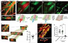
          Flow of cerebrospinal fluid (CSF) through perivascular spaces (PVSs) in the brain is important for clearance of metabolic waste. Arterial pulsations are thought to drive flow, but this has never been quantitatively shown. We used particle tracking to quantify CSF flow velocities in PVSs of live mice. CSF flow is pulsatile and driven primarily by the cardiac cycle. The speed of the arterial wall matches that of the CSF, suggesting arterial wall motion is the principal driving mechanism, via a process known as perivascular pumping. Increasing blood pressure leaves the artery diameter unchanged but changes the pulsations of the arterial wall, increasing backflow and thereby reducing net flow in the PVS. Perfusion-fixation alters the normal flow direction and causes a 10-fold reduction in PVS size. We conclude that particle tracking velocimetry enables the study of CSF flow in unprecedented detail and that studying the PVS in vivo avoids fixation artifacts.

          Abstract

          Arterial pulsations are thought to drive CSF flow through perivascular spaces (PVSs), but this has never been quantitatively shown. Using particle tracking to quantify CSF flow velocities in PVSs of live mice, the authors show that flow speeds match the instantaneous speeds of the pulsing artery walls that form the inner boundaries of the PVSs.

          Related collections

          Most cited references40

          • Record: found
          • Abstract: found
          • Article: not found

          A paravascular pathway facilitates CSF flow through the brain parenchyma and the clearance of interstitial solutes, including amyloid β.

          Because it lacks a lymphatic circulation, the brain must clear extracellular proteins by an alternative mechanism. The cerebrospinal fluid (CSF) functions as a sink for brain extracellular solutes, but it is not clear how solutes from the brain interstitium move from the parenchyma to the CSF. We demonstrate that a substantial portion of subarachnoid CSF cycles through the brain interstitial space. On the basis of in vivo two-photon imaging of small fluorescent tracers, we showed that CSF enters the parenchyma along paravascular spaces that surround penetrating arteries and that brain interstitial fluid is cleared along paravenous drainage pathways. Animals lacking the water channel aquaporin-4 (AQP4) in astrocytes exhibit slowed CSF influx through this system and a ~70% reduction in interstitial solute clearance, suggesting that the bulk fluid flow between these anatomical influx and efflux routes is supported by astrocytic water transport. Fluorescent-tagged amyloid β, a peptide thought to be pathogenic in Alzheimer's disease, was transported along this route, and deletion of the Aqp4 gene suppressed the clearance of soluble amyloid β, suggesting that this pathway may remove amyloid β from the central nervous system. Clearance through paravenous flow may also regulate extracellular levels of proteins involved with neurodegenerative conditions, its impairment perhaps contributing to the mis-accumulation of soluble proteins.
            Bookmark
            • Record: found
            • Abstract: not found
            • Article: not found

            2017 ACC/AHA/AAPA/ABC/ACPM/AGS/APhA/ASH/ASPC/NMA/PCNA Guideline for the Prevention, Detection, Evaluation, and Management of High Blood Pressure in Adults

              Bookmark
              • Record: found
              • Abstract: found
              • Article: not found

              Brain-wide pathway for waste clearance captured by contrast-enhanced MRI.

              The glymphatic system is a recently defined brain-wide paravascular pathway for cerebrospinal fluid (CSF) and interstitial fluid (ISF) exchange that facilitates efficient clearance of solutes and waste from the brain. CSF enters the brain along para-arterial channels to exchange with ISF, which is in turn cleared from the brain along para-venous pathways. Because soluble amyloid β clearance depends on glymphatic pathway function, we proposed that failure of this clearance system contributes to amyloid plaque deposition and Alzheimer's disease progression. Here we provide proof of concept that glymphatic pathway function can be measured using a clinically relevant imaging technique. Dynamic contrast-enhanced MRI was used to visualize CSF-ISF exchange across the rat brain following intrathecal paramagnetic contrast agent administration. Key features of glymphatic pathway function were confirmed, including visualization of para-arterial CSF influx and molecular size-dependent CSF-ISF exchange. Whole-brain imaging allowed the identification of two key influx nodes at the pituitary and pineal gland recesses, while dynamic MRI permitted the definition of simple kinetic parameters to characterize glymphatic CSF-ISF exchange and solute clearance from the brain. We propose that this MRI approach may provide the basis for a wholly new strategy to evaluate Alzheimer's disease susceptibility and progression in the live human brain.
                Bookmark

                Author and article information

                Contributors
                d.h.kelley@rochester.edu
                Journal
                Nat Commun
                Nat Commun
                Nature Communications
                Nature Publishing Group UK (London )
                2041-1723
                19 November 2018
                19 November 2018
                2018
                : 9
                : 4878
                Affiliations
                [1 ]ISNI 0000 0004 1936 9166, GRID grid.412750.5, Center for Translational Neuromedicine, , University of Rochester Medical Center, ; Rochester, NY 14642 USA
                [2 ]ISNI 0000 0004 1936 9166, GRID grid.412750.5, Department of Neuroscience, , University of Rochester Medical Center, ; Rochester, NY 14642 USA
                [3 ]ISNI 0000 0004 1936 9174, GRID grid.16416.34, Department of Mechanical Engineering, , University of Rochester, ; Rochester, NY 14627 USA
                [4 ]ISNI 0000 0000 9678 1884, GRID grid.412449.e, China Medical University, ; Shenyang, 110122 China
                [5 ]ISNI 0000 0001 0674 042X, GRID grid.5254.6, Center for Translational Neuromedicine, Faculty of Health and Medical Sciences, , University of Copenhagen, ; 2200 Copenhagen, Denmark
                Author information
                http://orcid.org/0000-0001-5876-5397
                http://orcid.org/0000-0003-2083-0901
                http://orcid.org/0000-0001-9658-2954
                Article
                7318
                10.1038/s41467-018-07318-3
                6242982
                30451853
                d1d279b2-2862-4029-a576-2d89cad092bb
                © The Author(s) 2018

                Open Access This article is licensed under a Creative Commons Attribution 4.0 International License, which permits use, sharing, adaptation, distribution and reproduction in any medium or format, as long as you give appropriate credit to the original author(s) and the source, provide a link to the Creative Commons license, and indicate if changes were made. The images or other third party material in this article are included in the article’s Creative Commons license, unless indicated otherwise in a credit line to the material. If material is not included in the article’s Creative Commons license and your intended use is not permitted by statutory regulation or exceeds the permitted use, you will need to obtain permission directly from the copyright holder. To view a copy of this license, visit http://creativecommons.org/licenses/by/4.0/.

                History
                : 7 May 2018
                : 26 October 2018
                Categories
                Article
                Custom metadata
                © The Author(s) 2018

                Uncategorized
                Uncategorized

                Comments

                Comment on this article