8
views
0
recommends
+1 Recommend
0 collections
    0
    shares
      • Record: found
      • Abstract: found
      • Article: found
      Is Open Access

      Dermatoscopy and Optical Coherence Tomography in Vulvar High-Grade Squamous Intraepithelial Lesions and Lichen Sclerosus: A Prospective Observational Trial

      research-article

      Read this article at

          There is no author summary for this article yet. Authors can add summaries to their articles on ScienceOpen to make them more accessible to a non-specialist audience.

          Abstract

          This feasibility study presents dermatoscopy and dynamic optical coherence tomography as easy-to-use, well-tolerated and noninvasive imaging tools aiding recognition of vulvar high-grade intraepithelial lesions and lichen sclerosus.

          Objective

          This study aimed to examine potential discriminatory characteristics of dermatoscopy and dynamic optical coherence tomography (D-OCT) on vulvar high-grade squamous intraepithelial lesions (vHSIL) and lichen sclerosus (LS) compared with healthy vulvar skin.

          Methods

          A prospective observational clinical trial was performed in 10 healthy volunteers, 5 vHSIL and 10 LS patients. Noninvasive imaging measurements using dermatoscopy and D-OCT were obtained at several time points, including lesional and nonlesional vulvar skin. Morphologic features of vHSIL and LS were compared with healthy controls. Epidermal thickness and blood flow were determined using D-OCT. Patients reported tolerability of each study procedure, including reference vulvar biopsies. The main outcome measures were feasibility and tolerability of imaging modalities, dermatoscopy and OCT characteristics, OCT epidermal thickness and D-OCT dermal blood flow.

          Results

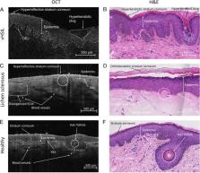
          The application of dermatoscopy and D-OCT is feasible and tolerable. In vHSIL, dermatoscopic warty structures were present. In LS, sclerotic areas and arborizing vessels were observed. Structural OCT in the vulvar area aligned with histology for hyperkeratosis and dermal-epidermal junction visualization. Currently, the OCT algorithm is unable to calculate the epidermal thickness of the uneven vulvar area. Dynamic optical coherence tomography showed statistically significant increased blood flow in LS patients (mean ± SD, 0.053 ± 0.029) to healthy controls (0.040 ± 0.012; p = .0024).

          Conclusions

          The application of dermatoscopy and D-OCT is feasible and tolerable in vHSIL and LS patients. Using dermatoscopy and D-OCT, the authors describe potential characteristics to aid differentiation of diseased from healthy vulvar skin, which could complement clinical assessments.

          Related collections

          Most cited references31

          • Record: found
          • Abstract: found
          • Article: found
          Is Open Access

          The Development, Commercialization, and Impact of Optical Coherence Tomography

          This review was written for the special issue of IOVS to describe the history of optical coherence tomography (OCT) and its evolution from a nonscientific, historic perspective. Optical coherence tomography has become a standard of care in ophthalmology, providing real-time information on structure and function – diagnosing disease, evaluating progression, and assessing response to therapy, as well as helping to understand disease pathogenesis and create new therapies. Optical coherence tomography also has applications in multiple clinical specialties, fundamental research, and manufacturing. We review the early history of OCT describing how research and development evolves and the important role of multidisciplinary collaboration and expertise. Optical coherence tomography had its origin in femtosecond optics, but used optical communications technologies and required advanced engineering for early OCT prototypes, clinical feasibility studies, entrepreneurship, and corporate development in order to achieve clinical acceptance and clinical impact. Critical advances were made by early career researchers, clinician scientists, engineering experts, and business leaders, which enabled OCT to have a worldwide impact on health care. We introduce the concept of an “ecosystem” consisting of research, government funding, collaboration and competition, clinical studies, innovation, entrepreneurship and industry, and impact – all of which must work synergistically. The process that we recount is long and challenging, but it is our hope that it might inspire early career professionals in science, engineering, and medicine, and that the clinical and research community will find this review of interest.
            Bookmark
            • Record: found
            • Abstract: found
            • Article: not found

            Squamous precursor lesions of the vulva: current classification and diagnostic challenges.

            Growing evidence has established two major types of vulvar intraepithelial neoplasia (VIN), which correspond to two distinct oncogenic pathways to vulvar squamous cell carcinoma (VSCC). While the incidence of VSCC has remained relatively stable over the last three decades, the incidence of VIN has increased. VIN of usual type (uVIN) is human papillomavirus (HPV)-driven, affects younger women and is a multicentric disease. In contrast, VIN of differentiated type (dVIN) occurs in post-menopausal women and develops independent of HPV infection. dVIN often arises in a background of lichen sclerosus and chronic inflammatory dermatoses. Although isolated dVIN is significantly less common than uVIN, dVIN bears a greater risk for malignant transformation to VSCC and progresses over a shorter time interval. On histological examination, uVIN displays conspicuous architectural and cytological abnormalities, while the morphological features that characterise dVIN are much more subtle and raise a wide differential diagnosis. On the molecular level, dVIN is characterised by a higher number of somatic mutations, particularly in TP53. Here we review the classification, epidemiology, clinical features, histomorphology, ancillary markers and molecular genetics of both types of VIN, and discuss the morphological challenges faced by pathologists in interpreting these lesions.
              Bookmark
              • Record: found
              • Abstract: found
              • Article: found
              Is Open Access

              Applications and future directions for optical coherence tomography in dermatology*

                Bookmark

                Author and article information

                Contributors
                Journal
                J Low Genit Tract Dis
                J Low Genit Tract Dis
                JLGTD
                Journal of Lower Genital Tract Disease
                Lippincott Williams & Wilkins
                1089-2591
                1526-0976
                July 2023
                16 March 2023
                : 27
                : 3
                : 255-261
                Affiliations
                [1 ]Centre for Human Drug Research, Leiden, The Netherlands
                [2 ]Department of Obstetrics and Gynaecology, Leiden University Medical Center, Leiden, The Netherlands
                [3 ]Leiden Academic Centre for Drug Research, Leiden University, Leiden, The Netherlands
                [4 ]Department of Obstetrics and Gynaecology and Catharina Cancer Institute, Catharina Ziekenhuis, Eindhoven, The Netherlands
                [5 ]Department of Pathology, Erasmus MC, University Medical Center, Rotterdam, The Netherlands
                [6 ]Department of Dermatology, Erasmus Medical Centre, Rotterdam, The Netherlands
                Author notes
                [*]Correspondence to: Prof. Robert Rissmann, PhD, PharmD; CHDR, Dermatology, Zernikedreef 8, 2333CL, Leiden. E-mail: rrissmannn@ 123456chdr.nl
                Article
                JLGTD_230142 00014
                10.1097/LGT.0000000000000731
                10309090
                36924426
                7c651c61-f5fb-4674-8ef4-0d905159b1fa
                Copyright © 2023 The Author(s). Published by Wolters Kluwer Health, Inc. on behalf of the ASCCP.

                This is an open access article distributed under the Creative Commons Attribution License 4.0 (CCBY), which permits unrestricted use, distribution, and reproduction in any medium, provided the original work is properly cited.

                History
                Categories
                HPV Associated Anal Disease
                Custom metadata
                TRUE
                T

                dermatoscopy,d-oct,lichen sclerosus,vulvar hsil
                dermatoscopy, d-oct, lichen sclerosus, vulvar hsil

                Comments

                Comment on this article