3
views
0
recommends
+1 Recommend
0 collections
    0
    shares
      • Record: found
      • Abstract: found
      • Article: found
      Is Open Access

      Contrast-enhanced mammography: what the radiologist needs to know

      review-article

      Read this article at

          There is no author summary for this article yet. Authors can add summaries to their articles on ScienceOpen to make them more accessible to a non-specialist audience.

          Abstract

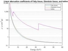
          Contrast-enhanced mammography (CEM) is a combination of standard mammography and iodinated contrast material administration. During the last decade, CEM has found its place in breast imaging protocols: after i.v. administration of iodinated contrast material, low-energy and high-energy images are retrieved in one acquisition using a dual-energy technique, and a recombined image is constructed enabling visualisation of areas of contrast uptake.

          The increased incorporation of CEM into everyday clinical practice is reflected in the installation of dedicated equipment worldwide, the (commercial) availability of systems from different vendors, the number of CEM examinations performed, and the number of scientific articles published on the subject. It follows that ever more radiologists will be confronted with this technique, and thus be required to keep up to date with the latest developments in the field. Most importantly, radiologists must have sufficient knowledge on how to interpret CEM images and be acquainted with common artefacts and pitfalls.

          This comprehensive review provides a practical overview of CEM technique, including CEM-guided biopsy; reading, interpretation and structured reporting of CEM images, including the accompanying learning curve, CEM artefacts and interpretation pitfalls; indications for CEM; disadvantages of CEM; and future developments.

          Related collections

          Most cited references79

          • Record: found
          • Abstract: found
          • Article: not found

          Mammographic density and the risk and detection of breast cancer.

          Extensive mammographic density is associated with an increased risk of breast cancer and makes the detection of cancer by mammography difficult, but the influence of density on risk according to method of cancer detection is unknown. We carried out three nested case-control studies in screened populations with 1112 matched case-control pairs. We examined the association of the measured percentage of density in the baseline mammogram with risk of breast cancer, according to method of cancer detection, time since the initiation of screening, and age. As compared with women with density in less than 10% of the mammogram, women with density in 75% or more had an increased risk of breast cancer (odds ratio, 4.7; 95% confidence interval [CI], 3.0 to 7.4), whether detected by screening (odds ratio, 3.5; 95% CI, 2.0 to 6.2) or less than 12 months after a negative screening examination (odds ratio, 17.8; 95% CI, 4.8 to 65.9). Increased risk of breast cancer, whether detected by screening or other means, persisted for at least 8 years after study entry and was greater in younger than in older women. For women younger than the median age of 56 years, 26% of all breast cancers and 50% of cancers detected less than 12 months after a negative screening test were attributable to density in 50% or more of the mammogram. Extensive mammographic density is strongly associated with the risk of breast cancer detected by screening or between screening tests. A substantial fraction of breast cancers can be attributed to this risk factor. Copyright 2007 Massachusetts Medical Society.
            Bookmark
            • Record: found
            • Abstract: found
            • Article: not found

            Breast MRI: State of the Art

            MRI of the breast has the highest sensitivity for breast cancer detection among current clinical imaging modalities and is indispensable for breast imaging practice. While the basis of breast MRI consists of T1-weighted contrast-enhanced imaging, T2-weighted, ultrafast, and diffusion-weighted imaging may be used to improve lesion characterization. Such multiparametric assessment of breast lesions allows for excellent discrimination between benign and malignant breast lesions. Indications for breast MRI are expanding. In preoperative staging, multiple studies confirm the superiority of MRI to other imaging modalities for tumor size estimation and detection of additional tumor foci in the ipsilateral and contralateral breast. Ongoing studies show that in experienced hands this can be used to improve breast cancer surgery, although there is no evidence of improved long-term outcomes. Screening indications are likewise growing as evidence is accumulating that OncologicRI depicts cancers at an earlier stage than mammography in all women. To manage the associated costs for screening, the use of abbreviated protocols may be beneficial. In patients treated with neoadjuvant chemotherapy, MRI is used to document response. It is essential to realize that oncologic and surgical response are different, and evaluation should be adapted to the underlying question.
              Bookmark
              • Record: found
              • Abstract: found
              • Article: found
              Is Open Access

              An overview of mammographic density and its association with breast cancer

              In 2017, breast cancer became the most commonly diagnosed cancer among women in the US. After lung cancer, breast cancer is the leading cause of cancer-related mortality in women. The breast consists of several components, including milk storage glands, milk ducts made of epithelial cells, adipose tissue, and stromal tissue. Mammographic density (MD) is based on the proportion of stromal, epithelial, and adipose tissue. Women with high MD have more stromal and epithelial cells and less fatty adipose tissue, and are more likely to develop breast cancer in their lifetime compared to women with low MD. Because of this correlation, high MD is an independent risk factor for breast cancer. Further, mammographic screening is less effective in detecting suspicious lesions in dense breast tissue, which can lead to late-stage diagnosis. Molecular differences between dense and non-dense breast tissues explain the underlying biological reasons for why women with dense breasts are at a higher risk for developing breast cancer. The goal of this review is to highlight the current molecular understanding of MD, its association with breast cancer risk, the demographics pertaining to MD, and the environmental factors that modulate MD. Finally, we will review the current legislation regarding the disclosure of MD on a traditional screening mammogram and the supplemental screening options available to women with dense breast tissue.
                Bookmark

                Author and article information

                Contributors
                Journal
                BJR Open
                BJR Open
                bjro
                BJR Open
                The British Institute of Radiology.
                2513-9878
                2021
                24 November 2021
                : 3
                : 1
                : 20210034
                Affiliations
                [1 ]org-divisionGROW School for Oncology and Developmental Biology, Maastricht University , Maastricht, the Netherlands
                [2 ]org-divisionDepartment of Radiology and Nuclear Medicine, Maastricht University Medical Center , Maastricht, the Netherlands
                [3 ]org-divisionDepartment of Medical Imaging, Laurentius Hospital , Roermond, the Netherlands
                [4 ]org-divisionDepartment of Radiology, Hospital del Mar , Barcelona, Spain
                [5 ]org-divisionDepartment of Surgery, Maastricht University Medical Center , Maastricht, the Netherlands
                [6 ]org-divisionDepartment of Medical Imaging, Zuyderland Medical Center , Sittard-Geleen, the Netherlands
                Author notes
                Address correspondence to: Lidewij M.F.H. Neeter. E-mail: lidewij.neeter@ 123456mumc.nl
                Author information
                https://orcid.org/0000-0003-2598-6219
                Article
                BJRO-D-21-00034
                10.1259/bjro.20210034
                8611680
                e4552014-bf44-4dc8-8e0a-033faeba7816
                © 2021 The Authors. Published by the British Institute of Radiology

                This is an open access article distributed under the terms of the Creative Commons Attribution 4.0 International License, which permits unrestricted use, distribution and reproduction in any medium, provided the original author and source are credited.

                History
                : 31 May 2021
                : 29 July 2021
                : 30 July 2021
                Page count
                Figures: 7, Tables: 3, Equations: 0, References: 83, Pages: 13, Words: 9273
                Categories
                Review Article
                bjro, BJRO
                brst, Breast

                Comments

                Comment on this article