11
views
0
recommends
+1 Recommend
0 collections
    0
    shares
      • Record: found
      • Abstract: found
      • Article: found
      Is Open Access

      Silicified collagen scaffold induces semaphorin 3A secretion by sensory nerves to improve in-situ bone regeneration

      research-article

      Read this article at
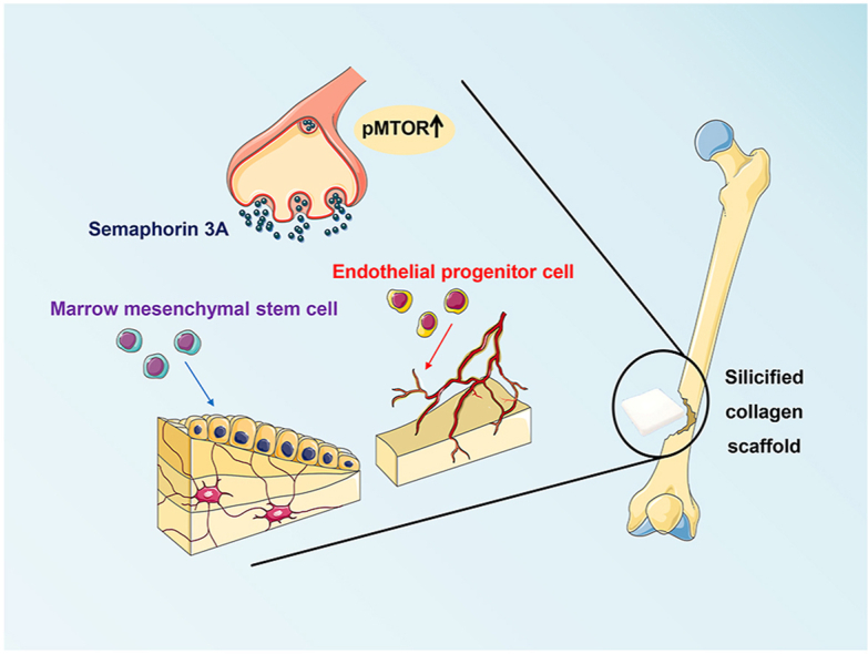
          There is no author summary for this article yet. Authors can add summaries to their articles on ScienceOpen to make them more accessible to a non-specialist audience.

          Abstract

          Sensory nerves promote osteogenesis through the release of neuropeptides. However, the potential application and mechanism in which sensory nerves promote healing of bone defects in the presence of biomaterials remain elusive. The present study identified that new bone formation was more abundantly produced after implantation of silicified collagen scaffolds into defects created in the distal femur of rats. The wound sites were accompanied by extensive nerve innervation and angiogenesis. Sensory nerve dysfunction by capsaicin injection resulted in significant inhibition of silicon-induced osteogenesis in the aforementioned rodent model. Application of extracellular silicon in vitro induced axon outgrowth and increased expression of semaphorin 3 A (Sema3A) and semaphorin 4D (Sema4D) in the dorsal root ganglion (DRG), as detected by the upregulation of signaling molecules. Culture medium derived from silicon-stimulated DRG cells promoted proliferation and differentiation of bone marrow mesenchymal stem cells and endothelial progenitor cells. These effects were inhibited by the use of Sema3A neutralizing antibodies but not by Sema4D neutralizing antibodies. Knockdown of Sema3A in DRG blocked silicon-induced osteogenesis and angiogenesis almost completely in a femoral defect rat model, whereas overexpression of Sema3A promoted the silicon-induced phenomena. Activation of “mechanistic target of rapamycin” (mTOR) pathway and increase of Sema3A production were identified in the DRG of rats that were implanted with silicified collagen scaffolds. These findings support the role of silicon in inducing Sema3A production by sensory nerves, which, in turn, stimulates osteogenesis and angiogenesis. Taken together, silicon has therapeutic potential in orthopedic rehabilitation.

          Graphical abstract

          Highlights

          • Nerve innervation, vascularization and tissue mineralization integrated into a single scaffold.

          • Silicified collagen scaffolds has therapeutic potential in orthopedic rehabilitation.

          • Silicified collagen scaffolds promote  in-situ bone regeneration via sensory nerve innervation and semaphorin 3A production.

          Related collections

          Most cited references58

          • Record: found
          • Abstract: found
          • Article: not found

          mTOR Signaling in Growth, Metabolism, and Disease.

          The mechanistic target of rapamycin (mTOR) coordinates eukaryotic cell growth and metabolism with environmental inputs, including nutrients and growth factors. Extensive research over the past two decades has established a central role for mTOR in regulating many fundamental cell processes, from protein synthesis to autophagy, and deregulated mTOR signaling is implicated in the progression of cancer and diabetes, as well as the aging process. Here, we review recent advances in our understanding of mTOR function, regulation, and importance in mammalian physiology. We also highlight how the mTOR signaling network contributes to human disease and discuss the current and future prospects for therapeutically targeting mTOR in the clinic.
            Bookmark
            • Record: found
            • Abstract: found
            • Article: not found

            Coupling of angiogenesis and osteogenesis by a specific vessel subtype in bone.

            The mammalian skeletal system harbours a hierarchical system of mesenchymal stem cells, osteoprogenitors and osteoblasts sustaining lifelong bone formation. Osteogenesis is indispensable for the homeostatic renewal of bone as well as regenerative fracture healing, but these processes frequently decline in ageing organisms, leading to loss of bone mass and increased fracture incidence. Evidence indicates that the growth of blood vessels in bone and osteogenesis are coupled, but relatively little is known about the underlying cellular and molecular mechanisms. Here we identify a new capillary subtype in the murine skeletal system with distinct morphological, molecular and functional properties. These vessels are found in specific locations, mediate growth of the bone vasculature, generate distinct metabolic and molecular microenvironments, maintain perivascular osteoprogenitors and couple angiogenesis to osteogenesis. The abundance of these vessels and associated osteoprogenitors was strongly reduced in bone from aged animals, and pharmacological reversal of this decline allowed the restoration of bone mass.
              Bookmark
              • Record: found
              • Abstract: found
              • Article: not found

              Leptin-receptor-expressing mesenchymal stromal cells represent the main source of bone formed by adult bone marrow.

              Studies of the identity and physiological function of mesenchymal stromal cells (MSCs) have been hampered by a lack of markers that permit both prospective identification and fate mapping in vivo. We found that Leptin Receptor (LepR) is a marker that highly enriches bone marrow MSCs. Approximately 0.3% of bone marrow cells were LepR(+), 10% of which were CFU-Fs, accounting for 94% of bone marrow CFU-Fs. LepR(+) cells formed bone, cartilage, and adipocytes in culture and upon transplantation in vivo. LepR(+) cells were Scf-GFP(+), Cxcl12-DsRed(high), and Nestin-GFP(low), markers which also highly enriched CFU-Fs, but negative for Nestin-CreER and NG2-CreER, markers which were unlikely to be found in CFU-Fs. Fate-mapping showed that LepR(+) cells arose postnatally and gave rise to most bone and adipocytes formed in adult bone marrow, including bone regenerated after irradiation or fracture. LepR(+) cells were quiescent, but they proliferated after injury. Therefore, LepR(+) cells are the major source of bone and adipocytes in adult bone marrow.
                Bookmark

                Author and article information

                Contributors
                Journal
                Bioact Mater
                Bioact Mater
                Bioactive Materials
                KeAi Publishing
                2452-199X
                27 July 2021
                March 2022
                27 July 2021
                : 9
                : 475-490
                Affiliations
                [a ]State Key Laboratory of Military Stomatology & National Clinical Research Center for Oral Diseases & Shaanxi Key Laboratory of Stomatology, School of Stomatology, The Fourth Military Medical University, Xi'an, Shaanxi, 710032, China
                [b ]Department of Neurobiology, The Fourth Military Medical University, Xi'an, Shaanxi, 710032, China
                [c ]The Graduate School, Augusta University, Augusta, GA, 30912, USA
                Author notes
                []Corresponding author. School of Stomatology, The Fourth Military Medical University, Xi'an, China. niulina831013@ 123456126.com niulina@ 123456fmmu.edu.cn
                [1]

                Equal contributors.

                Article
                S2452-199X(21)00352-2
                10.1016/j.bioactmat.2021.07.016
                8586786
                2f288bd8-77aa-41d6-9cda-ef591eddbde6
                © 2021 The Authors

                This is an open access article under the CC BY-NC-ND license (http://creativecommons.org/licenses/by-nc-nd/4.0/).

                History
                : 24 April 2021
                : 12 July 2021
                : 17 July 2021
                Categories
                Article

                distal femur defect,mechanistic target of rapamycin,semaphorin 3a,sensory nerve,silicified collagen scaffolds

                Comments

                Comment on this article