3
views
0
recommends
+1 Recommend
0 collections
    0
    shares
      • Record: found
      • Abstract: found
      • Article: found
      Is Open Access

      Characterization of the ExoU activation mechanism using EPR and integrative modeling

      research-article

      Read this article at

          There is no author summary for this article yet. Authors can add summaries to their articles on ScienceOpen to make them more accessible to a non-specialist audience.

          Abstract

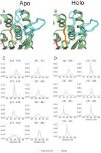
          ExoU, a type III secreted phospholipase effector of Pseudomonas aeruginosa, serves as a prototype to model large, dynamic, membrane-associated proteins. ExoU is synergistically activated by interactions with membrane lipids and ubiquitin. To dissect the activation mechanism, structural homology was used to identify an unstructured loop of approximately 20 residues in the ExoU amino acid sequence. Mutational analyses indicate the importance of specific loop amino acid residues in mediating catalytic activity. Engineered disulfide cross-links show that loop movement is required for activation. Site directed spin labeling EPR and DEER (double electron–electron resonance) studies of apo and holo states demonstrate local conformational changes at specific sites within the loop and a conformational shift of the loop during activation. These data are consistent with the formation of a substrate-binding pocket providing access to the catalytic site. DEER distance distributions were used as constraints in RosettaDEER to construct ensemble models of the loop in both apo and holo states, significantly extending the range for modeling a conformationally dynamic loop.

          Related collections

          Most cited references58

          • Record: found
          • Abstract: found
          • Article: not found

          ROSETTA3: an object-oriented software suite for the simulation and design of macromolecules.

          We have recently completed a full re-architecturing of the ROSETTA molecular modeling program, generalizing and expanding its existing functionality. The new architecture enables the rapid prototyping of novel protocols by providing easy-to-use interfaces to powerful tools for molecular modeling. The source code of this rearchitecturing has been released as ROSETTA3 and is freely available for academic use. At the time of its release, it contained 470,000 lines of code. Counting currently unpublished protocols at the time of this writing, the source includes 1,285,000 lines. Its rapid growth is a testament to its ease of use. This chapter describes the requirements for our new architecture, justifies the design decisions, sketches out central classes, and highlights a few of the common tasks that the new software can perform. © 2011 Elsevier Inc. All rights reserved.
            Bookmark
            • Record: found
            • Abstract: not found
            • Article: not found

            THE DISTRIBUTION OF THE FLORA IN THE ALPINE ZONE.1

              Bookmark
              • Record: found
              • Abstract: found
              • Article: not found

              Protein structure prediction and analysis using the Robetta server.

              The Robetta server (http://robetta.bakerlab.org) provides automated tools for protein structure prediction and analysis. For structure prediction, sequences submitted to the server are parsed into putative domains and structural models are generated using either comparative modeling or de novo structure prediction methods. If a confident match to a protein of known structure is found using BLAST, PSI-BLAST, FFAS03 or 3D-Jury, it is used as a template for comparative modeling. If no match is found, structure predictions are made using the de novo Rosetta fragment insertion method. Experimental nuclear magnetic resonance (NMR) constraints data can also be submitted with a query sequence for RosettaNMR de novo structure determination. Other current capabilities include the prediction of the effects of mutations on protein-protein interactions using computational interface alanine scanning. The Rosetta protein design and protein-protein docking methodologies will soon be available through the server as well.
                Bookmark

                Author and article information

                Contributors
                frankd@mcw.edu
                Journal
                Sci Rep
                Sci Rep
                Scientific Reports
                Nature Publishing Group UK (London )
                2045-2322
                12 November 2020
                12 November 2020
                2020
                : 10
                : 19700
                Affiliations
                [1 ]GRID grid.34477.33, ISNI 0000000122986657, Department of Chemistry, , University of Washington, ; Seattle, WA USA
                [2 ]GRID grid.30760.32, ISNI 0000 0001 2111 8460, Department of Microbiology and Immunology, , Medical College of Wisconsin, ; Milwaukee, WI USA
                [3 ]GRID grid.152326.1, ISNI 0000 0001 2264 7217, Department of Chemistry and Center for Structural Biology, , Vanderbilt University, ; Nashville, TN USA
                [4 ]GRID grid.152326.1, ISNI 0000 0001 2264 7217, Department of Molecular Physiology and Biophysics, , Vanderbilt University, ; Nashville, TN USA
                [5 ]GRID grid.10423.34, ISNI 0000 0000 9529 9877, Institute for Drug Discovery, , Leipzig University Medical School, ; Leipzig SAC, Germany
                [6 ]GRID grid.30760.32, ISNI 0000 0001 2111 8460, Department of Biophysics, , Medical College of Wisconsin, ; Milwaukee, WI USA
                Article
                76023
                10.1038/s41598-020-76023-3
                7665212
                33184362
                e6457742-75eb-464c-a2af-b0bf10609929
                © The Author(s) 2020

                Open Access This article is licensed under a Creative Commons Attribution 4.0 International License, which permits use, sharing, adaptation, distribution and reproduction in any medium or format, as long as you give appropriate credit to the original author(s) and the source, provide a link to the Creative Commons licence, and indicate if changes were made. The images or other third party material in this article are included in the article's Creative Commons licence, unless indicated otherwise in a credit line to the material. If material is not included in the article's Creative Commons licence and your intended use is not permitted by statutory regulation or exceeds the permitted use, you will need to obtain permission directly from the copyright holder. To view a copy of this licence, visit http://creativecommons.org/licenses/by/4.0/.

                History
                : 18 May 2020
                : 19 October 2020
                Funding
                Funded by: FundRef http://dx.doi.org/10.13039/100000002, National Institutes of Health;
                Award ID: AI104922
                Award ID: GM080403
                Categories
                Article
                Custom metadata
                © The Author(s) 2020

                Uncategorized
                biochemistry,biophysics,computational biology and bioinformatics
                Uncategorized
                biochemistry, biophysics, computational biology and bioinformatics

                Comments

                Comment on this article