2
views
0
recommends
+1 Recommend
0 collections
    0
    shares
      • Record: found
      • Abstract: found
      • Article: found
      Is Open Access

      Right Atrial Thrombus Presenting as Platypnea-Orthodeoxia Secondary to Reverse Lutembacher Syndrome: A Case Report

      case-report

      Read this article at

          There is no author summary for this article yet. Authors can add summaries to their articles on ScienceOpen to make them more accessible to a non-specialist audience.

          Abstract

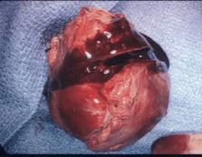
          Platypnea-orthodeoxia syndrome (POS) is defined by dyspnea and deoxygenation due to a change in body position from lying down to an upright position. We present a case of a large right atrial (RA) thrombus likely due to a right coronary artery fistula in a patient with a patent foramen ovale (PFO). On imaging, the thrombus was thought to be an atrial myxoma involving the tricuspid valve; however, after surgical excision and histopathological analysis, it was noted to be a cystic thrombus. Red-brown material along with vascular elements was noted on histopathology. Post-surgery, the patient was critically ill and died due to severe tricuspid regurgitation (TR) and hypotension despite using a right ventricle assist device and multiple vasopressors. Reverse Lutembacher syndrome (RLS) is defined as a triad of tricuspid stenosis (TS), elevated RA pressure, and right-to-left atrial shunting. The location of the mass and positional changes could be causing transient RLS from positional TS and interatrial shunting via the PFO causing POS. Cardiac magnetic resonance imaging can help differentiate between intracardiac masses. T1 and T2 signal characteristics and differences in contrast enhancement can help differentiate between a thrombus and a tumor. Treatment options include anticoagulation, thrombolysis, and thrombectomy. If severe TR occurs after surgery, treatment modalities such as caval valves could be an option in the future. Extracorporeal membrane oxygenation to provide right ventricle support in such cases could be considered.

          Related collections

          Most cited references21

          • Record: found
          • Abstract: found
          • Article: not found

          Clinical presentation of left atrial cardiac myxoma. A series of 112 consecutive cases.

          We analyzed a series of 112 consecutive cases of left atrial myxoma diagnosed in a single French hospital (72 women and 40 men; age range, 5-84 yr) over 40 years, from 1959 to 1998. Symptoms of mitral valve obstruction, the first arm of the classic triad of myxoma presentation, were present in 75 patients (67%), with mostly cardiac failure or malaise. Symptoms of embolism, the second frequent presentation in the classic triad, were observed in 33 cases (29%) with 1 or several locations, essentially cerebral emboli with stroke. Males are statistically at greater risk than females of developing embolic complications. The third arm of the classic triad consists of constitutional symptoms (34%) with fever, weight loss, or symptoms resembling connective tissue disease, due to cytokine (interleukin-6) secretion. Younger and male patients have more neurologic symptoms, and female patients have more systemic symptoms. Seventy-two patients (64%) had cardiac auscultation abnormalities, essentially pseudo-mitral valve disease (53.5%) and more rarely the suggestive tumor plop (15%). The most frequent electrocardiographic sign was left atrial hypertrophy (35%), whereas arrhythmias were uncommon. The greater number of myxoma patients (98) diagnosed preoperatively after 1977 reflects the introduction of echocardiography as a noninvasive diagnostic procedure. However, there was no significant reduction in the average time from onset of symptoms to operation between patients seen in the periods before and after 1977. The tumor diameter ranged from 1 to 15 cm with a weight of between 15 and 180 g (mean, 37 g). The myxoma surface was friable or villous in 35% of the cases, and smooth in the other 65% cases. Myxomas in patients presenting with embolism have a friable surface; those in patients with cardiac symptoms, pseudo-mitral auscultation signs, tumor plop, and electrocardiogram or radiologic signs of left atrium hypertrophy and dilatation are significantly the larger tumors. The long-term prognosis is excellent, and only 4 deaths occurred among our 112 cases over a median follow-up of 3 years. The recurrence rate is low (5%), but long-term follow-up and serial echocardiography are advisable especially for young patients.
            Bookmark
            • Record: found
            • Abstract: found
            • Article: not found

            The multiple dimensions of Platypnea-Orthodeoxia syndrome: A review

            Platypnea-Orthodeoxia syndrome (POS) is a rare clinical entity characterized by dyspnea and arterial desaturation while in the upright position. The various pathophysiologic mechanisms leading to POS has puzzled clinicians for years. The hypoxia in POS has been attributed to the mixing of the deoxygenated venous blood with the oxygenated arterial blood via a shunt. The primary mechanisms of POS in these patients can be broadly classified based on intracardiac abnormalities, extracardiac abnormalities and miscellaneous etiologies. A Patent Foramen Ovale (PFO) was the most common reported site of an intracardiac shunt. In addition to PFO, intracardiac shunt leading to POS has been reported from either an Atrial Septal Defect (ASD) or an Atrial Septal Aneurysm (ASA). Most patients with an intracardiac shunt also demonstrated a secondary anatomic or a functional defect. Extracardiac causes of POS included intra-pulmonary arteriovenous malformations and lung parenchymal diseases. A systematic evaluation is necessary to identify the underlying cause and institute an appropriate intervention. We conducted a review of literature and reviewed 239 cases of POS. In this article, we review the etiology and pathophysiology of POS and also summarize the diagnostic algorithms and treatment modalities available for early diagnosis and prompt treatment of patients presenting with symptoms of platypnea and/or orthodeoxia.
              Bookmark
              • Record: found
              • Abstract: not found
              • Article: not found

              Platypnea-orthodeoxia syndrome: etiology, differential diagnosis, and management.

              T O Cheng (1999)
                Bookmark

                Author and article information

                Journal
                Cureus
                Cureus
                2168-8184
                Cureus
                Cureus (Palo Alto (CA) )
                2168-8184
                11 July 2022
                July 2022
                : 14
                : 7
                : e26754
                Affiliations
                [1 ] Internal Medicine, University of South Dakota Sanford School of Medicine, Sioux Falls, USA
                [2 ] Pathology, University of South Dakota Sanford School of Medicine, Sioux Falls, USA
                [3 ] Pulmonary and Critical Care Medicine, Sanford University of South Dakota (USD) Medical Center, Sioux Falls, USA
                Author notes
                Article
                10.7759/cureus.26754
                9275549
                35836710
                e2fc4650-e147-4977-be0b-5eb02b633bf8
                Copyright © 2022, Hamid et al.

                This is an open access article distributed under the terms of the Creative Commons Attribution License, which permits unrestricted use, distribution, and reproduction in any medium, provided the original author and source are credited.

                History
                : 11 July 2022
                Categories
                Cardiology
                Pathology
                Pulmonology

                patent foramen ovale,impella device,tricuspid valve stenosis,coronary arterial fistula,platypnea-orthodeoxia syndrome,atrium thrombus,right atrial myxoma

                Comments

                Comment on this article