19
views
0
recommends
+1 Recommend
0 collections
    0
    shares
      • Record: found
      • Abstract: found
      • Article: found
      Is Open Access

      Ventriculostomy-related infections in subarachnoid hemorrhage patients—a retrospective study of incidence, etiology, and antimicrobial therapy

      research-article

      Read this article at

          There is no author summary for this article yet. Authors can add summaries to their articles on ScienceOpen to make them more accessible to a non-specialist audience.

          Abstract

          Background

          This study was performed to investigate the incidence and etiology of ventriculostomy-related infections (VRIs) in patients with subarachnoid hemorrhage (SAH) and to assess adherence to local clinical guidelines regarding empirical antimicrobial therapy and diagnostic routines.

          Methods

          A total of 191 consecutive SAH patients treated in the neuro-intensive care unit of Uppsala University Hospital between 2010 and 2013 were included retrospectively. Information regarding cerebrospinal fluid samples, bacterial cultures, ventriculostomy treatment, patient characteristics, and antibiotic treatment were collected from electronic patient records.

          Results

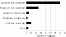
          Eleven patients developed VRI, resulting in an incidence of 5.8% per patient, 5.4% per ventriculostomy catheter, and 4.1 per 1000 catheter days. Coagulase-negative staphylococci caused nine cases of VRI and Klebsiella pneumoniae and Staphylococcus aureus caused one each. Empirical VRI therapy was initiated on 97 occasions in 81 subjects (42.4%). Out of the 11 patients with VRI, four did not receive empirical antibiotic therapy before the positive culture result. The clinical actions performed after analysis of CSF samples were in line with the action suggested by the local guidelines in 307 out of 592 cases (51.9%).

          Conclusions

          The incidence of VRI in our cohort was comparable to what has previously been reported. Coagulase-negative staphylococci was the most common agent. Our study demonstrates the difficulty in diagnosing VRI in SAH patients. Improved adherence to clinical guidelines could to some extent reduce the use of empirical antibiotic treatment, but better diagnostic methods and routines are needed.

          Related collections

          Most cited references36

          • Record: found
          • Abstract: found
          • Article: not found

          Coagulase-negative staphylococci.

          The definition of the heterogeneous group of coagulase-negative staphylococci (CoNS) is still based on diagnostic procedures that fulfill the clinical need to differentiate between Staphylococcus aureus and those staphylococci classified historically as being less or nonpathogenic. Due to patient- and procedure-related changes, CoNS now represent one of the major nosocomial pathogens, with S. epidermidis and S. haemolyticus being the most significant species. They account substantially for foreign body-related infections and infections in preterm newborns. While S. saprophyticus has been associated with acute urethritis, S. lugdunensis has a unique status, in some aspects resembling S. aureus in causing infectious endocarditis. In addition to CoNS found as food-associated saprophytes, many other CoNS species colonize the skin and mucous membranes of humans and animals and are less frequently involved in clinically manifested infections. This blurred gradation in terms of pathogenicity is reflected by species- and strain-specific virulence factors and the development of different host-defending strategies. Clearly, CoNS possess fewer virulence properties than S. aureus, with a respectively different disease spectrum. In this regard, host susceptibility is much more important. Therapeutically, CoNS are challenging due to the large proportion of methicillin-resistant strains and increasing numbers of isolates with less susceptibility to glycopeptides.
            Bookmark
            • Record: found
            • Abstract: found
            • Article: not found

            Relation of cerebral vasospasm to subarachnoid hemorrhage visualized by computerized tomographic scanning.

            In 47 cases of verified ruptured saccular aneurysm, we investigated the relationship of the amount and distribution of subarachnoid blood detected by computerized tomography to the later development of cerebral vasospasm. When the subarachnoid blood was not detected or was distributed diffusely, severe vasospasm was almost never encounters (1 of 18 cases). In the presence of subarachnoid blood clots larger than 5 X 3 mm (measured on the reproduced images) or layers of blood 1 mm or more thick in fissures and vertical cisterns, severe spasm followed almost invariably (23 of 24 cases). There was an almost exact correspondence between the site of the major subarachnoid blood clots and the location of severe vasospasm. Every patient with severe vasospasm manifested delayed symptoms and signs. Excellent correlation existed between the particular artery in vasospasm and the delayed clinical syndrome. Severe vasospasm involved the anterior cerebral artery in 20 cases and the middle cerebral artery in only 14. As the grading system used is partly subjective, the findings should be regarded as preliminary. The results, if confirmed, indicate that blood localized in the subarachnoid space in sufficient amount at specific sites is the only important etiological factor in vasospasm. It should be possible to identify patients in jeopardy from vasospasm and institute early preventive measures. (Neurosurgery, 6: 1--9, 1980)
              Bookmark
              • Record: found
              • Abstract: found
              • Article: not found

              Case-fatality rates and functional outcome after subarachnoid hemorrhage: a systematic review.

              During the last three decades, new management strategies have been developed for patients with aneurysmal subarachnoid hemorrhage. To assess whether the case-fatality rate has improved after the introduction of new management strategies, we studied outcome in all population-based studies from 1960 onward. To identify population-based studies that reported on case-fatality rate in subarachnoid hemorrhage, we performed a MEDLINE search and checked all reference lists of the studies found. Two authors (J.W.H. and G.J.E.R.) independently assessed all studies for eligibility, using predefined criteria for case finding and diagnosis, and extracted data on case-fatality rates. We used weighted linear regression analysis to quantify change in case-fatality rate over time. We found 21 studies, describing 25 study periods between 1960 and 1992. Case-fatality rates varied between 32% and 67%, with the exception of one recent study. The case-fatality rate decreased by 0.5% per year (95% confidence interval, -0.1 to 1.2); the decline was steeper after adjustment for age and sex (0.9% per year; 95% confidence interval, -0.7 to 2.6; data from 12 studies). The case-fatality rate after subarachnoid hemorrhage has decreased during the last three decades. A plausible explanation for this decrease is the improved management of patients with subarachnoid hemorrhage.
                Bookmark

                Author and article information

                Contributors
                +4618 611 56 40 , gabriel.westman@medsci.uu.se
                Journal
                Acta Neurochir (Wien)
                Acta Neurochir (Wien)
                Acta Neurochirurgica
                Springer Vienna (Vienna )
                0001-6268
                0942-0940
                7 December 2016
                7 December 2016
                2017
                : 159
                : 2
                : 317-323
                Affiliations
                [1 ]Department of Medical Sciences, Uppsala University, 751 85 Uppsala, Sweden
                [2 ]Department of Neuroscience, Uppsala University, Uppsala, Sweden
                Article
                3039
                10.1007/s00701-016-3039-2
                5241344
                27928632
                7337ebad-14dd-47d4-b41d-941a52b4684f
                © The Author(s) 2016

                Open Access This article is distributed under the terms of the Creative Commons Attribution 4.0 International License (http://creativecommons.org/licenses/by/4.0/), which permits unrestricted use, distribution, and reproduction in any medium, provided you give appropriate credit to the original author(s) and the source, provide a link to the Creative Commons license, and indicate if changes were made.

                History
                : 21 August 2016
                : 22 November 2016
                Funding
                Funded by: Uppsala Universitet (SE)
                Funded by: Akademiska Sjukhuset (SE)
                Categories
                Original Article - Vascular
                Custom metadata
                © Springer-Verlag Wien 2017

                Surgery
                ventriculostomy,meningitis,subarachnoid hemorrhage,antimicrobial therapy
                Surgery
                ventriculostomy, meningitis, subarachnoid hemorrhage, antimicrobial therapy

                Comments

                Comment on this article